Explore
 Validate
 Learn
Learn Western blot
 Immunocytochemistry
 Immunoprecipitation
Antibody data
- Antibody Data
- Antigen structure
- References [1]
- Comments [0]
- Validations
- Immunocytochemistry [2]
Submit
Validation data
Reference
Comment
Report error
- Product number
- MA5-27720 - Provider product page

- Provider
- Invitrogen Antibodies
- Product name
- ATP7A Monoclonal Antibody (L60/4)
- Antibody type
- Monoclonal
- Antigen
- Synthetic peptide
- Description
- 1 µg/mL of MA5-27720 was sufficient for detection of Copper-transporting ATPase1 in 20 µg of rat brain lysate by colorimetric immunoblot analysis using Goat IgG:HRP as the secondary antibody.|Detects approximately 180kDa in rat brain membrane preparations. This antibody was formerly sold as clone S60-4.
- Reactivity
- Human, Mouse, Rat
- Host
- Mouse
- Isotype
- IgG
- Antibody clone number
- L60/4
- Vial size
- 100 μg
- Concentration
- 1 mg/mL
- Storage
- -20°C
Submitted references Streamlined copper defenses make Bordetella pertussis reliant on custom-made operon.
Rivera-Millot A, Slupek S, Chatagnon J, Roy G, Saliou JM, Billon G, Alaimo V, Hot D, Salomé-Desnoulez S, Locht C, Antoine R, Jacob-Dubuisson F
Communications biology 2021 Jan 8;4(1):46
Communications biology 2021 Jan 8;4(1):46
No comments: Submit comment
Supportive validation
- Submitted by
- Invitrogen Antibodies (provider)
- Main image

- Experimental details
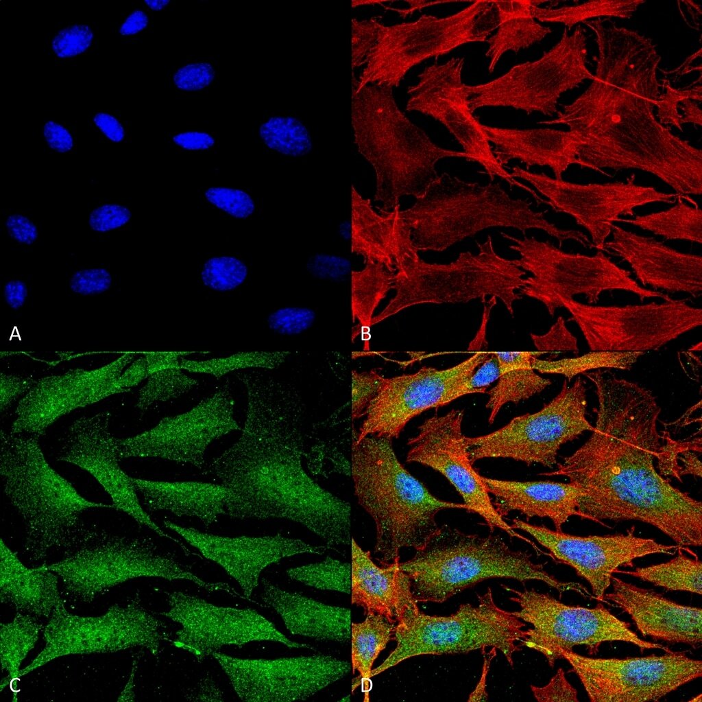
- Immunofluorescent analysis of ATP7A in mouse NIH 3T3 (mouse fibroblast cell line). Sample was fixed with 4% formaldehyde (15 min at RT), incubated with ATP7A monoclonal antibody (Product # MA5-27720) using a dilution of 1:100 (1 hour at RT), and followed by Goat Anti-Mouse 488, Phalloidin Texas Red and DAPI secondary antibody at a dilution of 1:200, 1:1000 (60 min at RT) and 1:5000 (5 min at RT). Images are shown as follows: (A) DAPI (blue) nuclear stain, B) Phalloidin Texas Red F-Actin stain, C) Copper Transporting ATPase 1 Antibody, D) Merged image. Magnification: 60x.

- Submitted by
- Invitrogen Antibodies (provider)
- Main image

- Experimental details
- Immunofluorescent analysis of ATP7A in mouse NIH 3T3 (mouse fibroblast cell line). Sample was fixed with 4% formaldehyde (15 min at RT), incubated with ATP7A monoclonal antibody (Product # MA5-27720) using a dilution of 1:100 (1 hour at RT), and followed by Goat Anti-Mouse 488, Phalloidin Texas Red and DAPI secondary antibody at a dilution of 1:200, 1:1000 (60 min at RT) and 1:5000 (5 min at RT). Images are shown as follows: (A) DAPI (blue) nuclear stain, B) Phalloidin Texas Red F-Actin stain, C) Copper Transporting ATPase 1 Antibody, D) Merged image. Magnification: 60x.