Explore
 Validate
 Learn
Learn Western blot
Antibody data
- Antibody Data
- Antigen structure
- References [0]
- Comments [0]
- Validations
- Western blot [1]
- ELISA [1]
- Other assay [3]
Submit
Validation data
Reference
Comment
Report error
- Product number
- TA347143 - Provider product page

- Provider
- OriGene
- Product name
- Rabbit Polyclonal H2BK12ac Antibody
- Antibody type
- Polyclonal
- Description
- Rabbit Polyclonal H2BK12ac Antibody
- Host
- Rabbit
- Conjugate
- Unconjugated
- Epitope
- HIST1H2BD
- Isotype
- IgG
- Antibody clone number
- NULL
- Vial size
- 50 µg
- Concentration
- 0.82 ?g/?l
No comments: Submit comment
Supportive validation
- Submitted by
- OriGene (provider)
- Main image
- Experimental details
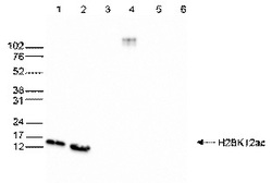
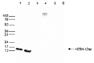
- WB was performed on whole cell (25 ug, lane 1) and histone extracts (15 ug, lane 2 ) from HeLa cells, and on 1 ug of recombinant histone H2A, H2B, H3 and H4 (lane 3, 4, 5 and 6, respectively) using the antibody against H2BK12ac. The antibody was diluted 1:1,000 in TBS-Tween containing 5% skimmed milk. The marker (in kDa) is shown on the left.
- Validation comment
- WB
Supportive validation
- Submitted by
- OriGene (provider)
- Main image
- Experimental details
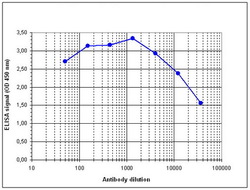
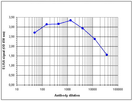
- Determination of the antibody titer To determine the titer of the antibody, an ELISA was performed using a serial dilution of the antibody against H2BK12ac in antigen coated wells. The antigen used was a peptide containing the histone modification of interest. By plotting the absorbance against the antibody dilution (Figure 3), the titer of the antibody was estimated to be 1:38,200.
- Validation comment
- ELISA
Supportive validation
- Submitted by
- OriGene (provider)
- Main image

- Experimental details
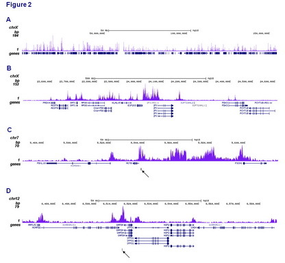
- ChIP was performed on sheared chromatin from 1.5 million HeLaS3 cells using 0.5 ug antibody. The 51 bp tags were aligned to the human genome using the BWA algorithm. Image shows the enrichment along the complete sequence and a 1Mb region of the X-chromosome (A and B) and in genomic regions of chromosome 7, surrounding ACTB gene, and of chromosome 12, surrounding GAPDH gene (C and D). The position of the amplicon used for ChIP-qPCR is indicated by arrow.
- Validation comment
- Assay
- Submitted by
- OriGene (provider)
- Main image
- Experimental details
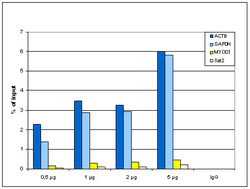
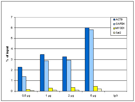
- ChIP assays using HeLa cells (sheared chromatin from 1.5 million cells). Titration of 0.5, 1, 2 and 5ug antibody per ChIP was analysed. IgG (1 ug/IP) was used as negative control. qPCR primers were for a region ~1kb upstream of GAPDH and ACTB promoters as positive controls, and for the coding region of inactive MYOD1 gene and Sat2 satellite repeat as negative controls. Image shows the recovery, expressed as a % of input (the relative amount of IP'd DNA compared to input DNA after qPCR).
- Validation comment
- Assay
- Submitted by
- OriGene (provider)
- Main image

- Experimental details
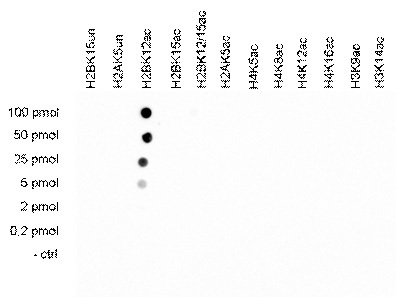
- A Dot Blot analysis was performed with peptides containing other histone modifications and the unmodified H2B. One hundred to 0.2 pmol of the respective peptides were spotted on a membrane. The antibody was used at a dilution of 1:5,000. Figure 4 shows a high specificity of the antibody for the modification of interest.
- Validation comment
- DB