 Explore
 Validate
 Learn
Learn Western blot
 ELISA
ELISA Immunohistochemistry
Antibody data
- Antibody Data
- Antigen structure
- References [0]
- Comments [0]
- Validations
- Western blot [1]
- Immunohistochemistry [2]
Submit
Validation data
Reference
Comment
Report error
- Product number
- LS-B10601 - Provider product page

- Provider
- LSBio
- Product name
- IHC-plus™ AHSA1 / AHA1 Antibody (clone 25F2.D9) LS-B10601
- Antibody type
- Monoclonal
- Description
- Protein G affinity chromatography
- Reactivity
- Human, Mouse
- Host
- Rat
- Isotype
- IgG
- Antibody clone number
- 25F2.D9
- Storage
- Short term: store at 4°C. Long term: aliquot and store at -20°C. Avoid freeze-thaw cycles.
No comments: Submit comment
Supportive validation
- Submitted by
- LSBio (provider)
- Enhanced method
- Genetic validation
- Main image

- Experimental details
- Anti-AHA1 Monoclonal Antibody - Western Blot. Western blot of anti-AHA1 monoclonal antibody shows detection of a band ~42 kD in size corresponding to AHA1 in A431 whole cell lysate (lane 1) and MCF-7 whole cell lysate (lane 2). A control lane is shown where primary ahntibody was omitted from the incubation (lane C). Molecular weight markers are shown at the left. For best results, block the membrane overnight with 3% BSA in TBS followed by reaction with primary antibody diluted 1:1000 and use HRP conjugated anti-Rabbit IgG (LS-C60865) secondary antibody diluted 1:20000 in blocking buffer (p/n MB-070) for detection.

Enhanced validation
- Submitted by
- LSBio (provider)
- Enhanced method
- Genetic validation
- Main image

- Experimental details
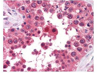
- Anti-AHA1 Monoclonal Antibody - Immunohistochemistry. anti-AHA1 monoclonal antibody was used at a 5-10 ug/mL to detect AHA1 in the seminiferous tubule of human testis (40X) showing moderate staining. Leydig cells showed faint to moderate staining. expression of AHA1 is reported in many epithelial and lymphatic tissues, with cytoplasmic localization. This antibody showed moderate cytoplasmic staining of a variety of epithelial tissues and lymphoid organs such as spleen and tonsil with minimal background staining. The image shows the localization of the antibody as the precipitated red signal, with a hematoxylin purple nuclear counterstain. Tissue was formalin-fixed and paraffin embedded.
- Submitted by
- LSBio (provider)
- Enhanced method
- Genetic validation
- Main image

- Experimental details
- Anti-AHA1 Monoclonal Antibody - Immunohistochemistry. anti-AHA1 monoclonal antibody was used at a 5-10 ug/mL to detect AHA1 in the seminiferous tubule of human testis (40X) showing moderate staining. Leydig cells showed faint to moderate staining. expression of AHA1 is reported in many epithelial and lymphatic tissues, with cytoplasmic localization. This antibody showed moderate cytoplasmic staining of a variety of epithelial tissues and lymphoid organs such as spleen and tonsil with minimal background staining. The image shows the localization of the antibody as the precipitated red signal, with a hematoxylin purple nuclear counterstain. Tissue was formalin-fixed and paraffin embedded.
