Explore
 Validate
 Learn
Learn Western blot
Antibody data
- Antibody Data
- Antigen structure
- References [3]
- Comments [0]
- Validations
- Western blot [1]
- Immunohistochemistry [1]
Submit
Validation data
Reference
Comment
Report error
- Product number
- MAB8169 - Provider product page

- Provider
- Abnova Corporation
- Proper citation
- Abnova Corporation Cat#MAB8169, RRID:AB_10678460
- Product name
- Ahsa1 monoclonal antibody, clone 25F2.D9
- Antibody type
- Monoclonal
- Description
- Rat monoclonal antibody raised against full length recombinant Ahsa1.
- Antibody clone number
- 25F2.D9
- Storage
- Store at 4°C. For long term storage store at -20°C.Aliquot to avoid repeated freezing and thawing.
Submitted references The charged linker region is an important regulator of Hsp90 function.
A novel HSP90 chaperone complex regulates intracellular vesicle transport.
Hsp90 cochaperone Aha1 downregulation rescues misfolding of CFTR in cystic fibrosis.
Hainzl O, Lapina MC, Buchner J, Richter K
The Journal of biological chemistry 2009 Aug 21;284(34):22559-67
The Journal of biological chemistry 2009 Aug 21;284(34):22559-67
A novel HSP90 chaperone complex regulates intracellular vesicle transport.
Lotz GP, Brychzy A, Heinz S, Obermann WM
Journal of cell science 2008 Mar 1;121(Pt 5):717-23
Journal of cell science 2008 Mar 1;121(Pt 5):717-23
Hsp90 cochaperone Aha1 downregulation rescues misfolding of CFTR in cystic fibrosis.
Wang X, Venable J, LaPointe P, Hutt DM, Koulov AV, Coppinger J, Gurkan C, Kellner W, Matteson J, Plutner H, Riordan JR, Kelly JW, Yates JR 3rd, Balch WE
Cell 2006 Nov 17;127(4):803-15
Cell 2006 Nov 17;127(4):803-15
No comments: Submit comment
Supportive validation
- Submitted by
- Abnova Corporation (provider)
- Main image
- Experimental details
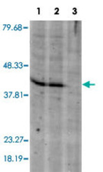
- Western blot using Ahsa1 monoclonal antibody, clone 25F2.D9 (Cat # MAB8169) shows detection of a band ~42 KDa in size corresponding to Ahsa1 in A-431 whole cell lysate (Lane 1) and MCF-7 whole cell lysate (Lane 2). A control lane is shown where primary ahntibody was omitted from the incubation (Lane 3). Molecular weight markers are shown at the left. For best results, block the membrane overnight with 3% BSA in TBS followed by reaction with primary antibody diluted 1 : 1,000 and use HRP conjugated anti-Rabbit IgG secondary antibody diluted 1 : 20,000 in blocking buffer for detection.

Supportive validation
- Submitted by
- Abnova Corporation (provider)
- Main image
- Experimental details
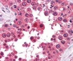
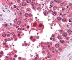
- Immunohistochemical staining of Ahsa1 monoclonal antibody, clone 25F2.D9 (Cat # MAB8169) was used at a 5-10 ug/mL to detect Ahsa1 in the seminiferous tubule of human testis (40X) showing moderate staining. Leydig cells showed faint to moderate staining. Expression of Ahsa1 is reported in many epithelial and lymphatic tissues, with cytoplasmic localization. This antibody showed moderate cytoplasmic staining of a variety of epithelial tissues and lymphoid organs such as spleen and tonsil with minimal background staining. The image shows the localization of the antibody as the precipitated red signal, with a hematoxylin purple nuclear counterstain. Tissue was formalin-fixed and paraffin embedded.Personal Communication, Vasiliki Demas, LifeSpanBiosciences, Seattle, WA.
- Validation comment
- Immunohistochemistry (Formalin/PFA-fixed paraffin-embedded sections)