 Explore
 Validate
 Learn
Learn Western blot
 Immunohistochemistry
Antibody data
- Antibody Data
- Antigen structure
- References [1]
- Comments [0]
- Validations
- Immunohistochemistry [1]
- Other assay [1]
Submit
Validation data
Reference
Comment
Report error
- Product number
- PA5-20076 - Provider product page

- Provider
- Invitrogen Antibodies
- Product name
- NADE Polyclonal Antibody
- Antibody type
- Polyclonal
- Antigen
- Synthetic peptide
- Description
- Despite its predicted molecular weight, NADE often migrates at ~23kDa in SDS-PAGE. A suggested positive control is human brain tissue lysate. PA5-20076 can be used with blocking peptide PEP-0194.
- Reactivity
- Human, Mouse
- Host
- Rabbit
- Isotype
- IgG
- Vial size
- 100 μg
- Concentration
- 1 mg/mL
- Storage
- Maintain refrigerated at 2-8°C for up to 3 months. For long term storage store at -20°C
Submitted references Saikosaponin-d impedes hippocampal neurogenesis and causes cognitive deficits by inhibiting the survival of neural stem/progenitor cells via neurotrophin receptor signaling in mice.
Qin T, Yuan Z, Yu J, Fu X, Deng X, Fu Q, Ma Z, Ma S
Clinical and translational medicine 2020 Dec;10(8):e243
Clinical and translational medicine 2020 Dec;10(8):e243
No comments: Submit comment
Supportive validation
- Submitted by
- Invitrogen Antibodies (provider)
- Main image
- Experimental details
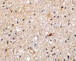
- Immunohistochemistry of NADE in human brain tissue with NADE Polyclonal Antibody (Product # PA5-20076) at 2 µg/mL.

Supportive validation
- Submitted by
- Invitrogen Antibodies (provider)
- Main image
- Experimental details
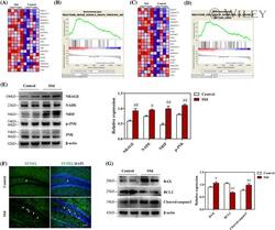
- 2 FIGURE SSd activated NRAGE/NADE/NRIF cell apoptotic signaling in the hippocampus. A, A heat map of the NRAGE pathway-related genes shows a strong correlation in SSd-treated mice (n = 5). B, GSEA profiles of the correlation between SSd-treated mice and NRAGE signaling. C, A heat map of the NRAGE/NRIF/NADE pathway-related genes shows a strong correlation in SSd-treated mice (n = 5). D, GSEA profiles of the correlation between SSd-treated mice and NRAGE/NRIF/NADE signaling. The upper part plots the enrichment scores for each gene, and the lower part of the plot shows the value of the ranking metric moving down the list of ranked genes. E, Western blot analysis of NRAGE/NRIF/NADE signaling in the hippocampus and quantitative analysis of the levels of NRAGE, NRIF, NADE, and p-JNK (n = 4). F, Hippocampal cell apoptosis was evaluated by TUNEL staining in the DG (n = 4). Scale bar 50 mum. G, Western blot analysis of the apoptotic proteins in the hippocampus and quantitative analysis of the levels of BAX, BCL2, cleaved caspase-3 (n = 4). Data are expressed as mean +- SEM. # P < .05 versus the control. ## P < .01 versus the control
