Explore
 Validate
 Learn
Learn Western blot
 Immunocytochemistry
Antibody data
- Antibody Data
- Antigen structure
- References [0]
- Comments [0]
- Validations
- Immunocytochemistry [2]
- Immunohistochemistry [1]
- Flow cytometry [2]
Submit
Validation data
Reference
Comment
Report error
- Product number
- PA5-72054 - Provider product page

- Provider
- Invitrogen Antibodies
- Product name
- Rubicon Polyclonal Antibody
- Antibody type
- Polyclonal
- Antigen
- Synthetic peptide
- Reactivity
- Human
- Host
- Rabbit
- Isotype
- IgG
- Vial size
- 400 μL
- Concentration
- 0.4 mg/mL
- Storage
- Store at 4°C short term. For long term storage, store at -20°C, avoiding freeze/thaw cycles.
No comments: Submit comment
Supportive validation
- Submitted by
- Invitrogen Antibodies (provider)
- Main image

- Experimental details
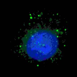
- Immunofluorescent analysis of BARON in U251 cells. Samples were treated with Chloroquine (50 µM,16h), then fixed with 4% paraformaldehyde (20 min), permeabilized with Triton X-100 (0.2%, 30 min). Cells were then incubated with PA5-72054 BARON (N-term) primary antibody (1:100, 2 h at room temperature). For secondary antibody, Alexa Fluor® 488 conjugated donkey anti-rabbit antibody (green) was used (1:1000, 1h). Nuclei were counterstained with Hoechst 33342 (blue) (10 µg/mL, 5 min).

- Submitted by
- Invitrogen Antibodies (provider)
- Main image
- Experimental details
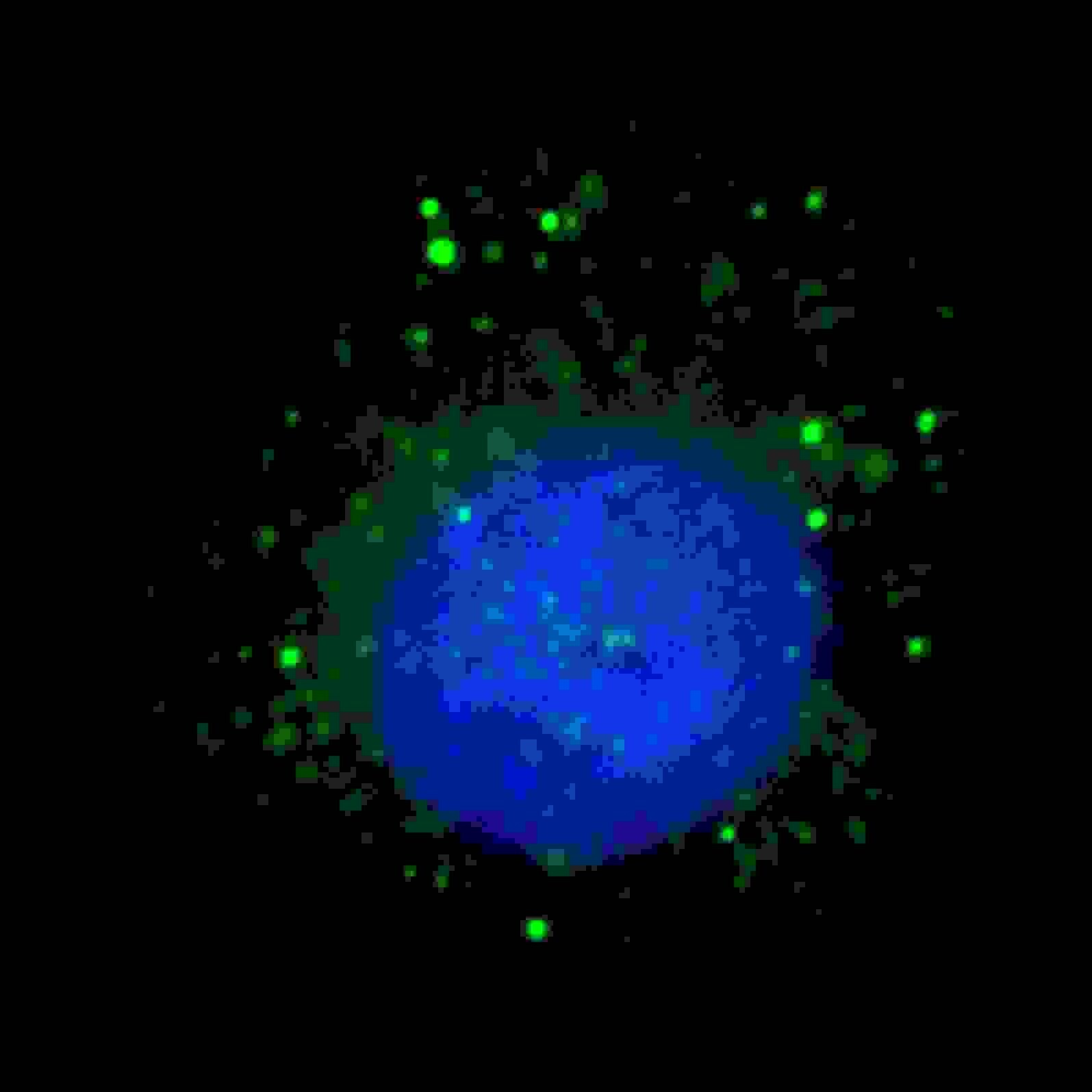
- Immunocytochemistry analysis of Rubicon in U251 cells. Samples were incubated with Rubicon polyclonal antibody (Product # PA5-72054) using a dilution of 1:100 for 2 hr at room temperature followed by Alexa Fluor® 488 conjugated donkey anti-rabbit antibody (green) at a dilution of 1:1,000 for 1hr. Cells were treated with Chloroquine (50 μM, 16hr), then fixed with 4% PFA (20 min), and permeabilized with Triton X-100 (0.2%, 30 min). Nuclei were counterstained with Hoechst 33342 (blue) (10 µg/mL, 5 min). BARON immunoreactivity is localized to autophagic vacuoles in the cytoplasm of U251 cells.
Supportive validation
- Submitted by
- Invitrogen Antibodies (provider)
- Main image
- Experimental details
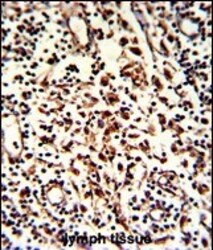
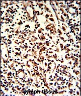
- Immunohistochemistry analysis of Rubicon in formalin-fixed and paraffin-embedded human lymph. Samples were incubated with Rubicon polyclonal antibody (Product # PA5-72054) which was peroxidase-conjugated to the secondary antibody, followed by DAB staining. This data demonstrates the use of this antibody for immunohistochemistry; clinical relevance has not been evaluated.
Supportive validation
- Submitted by
- Invitrogen Antibodies (provider)
- Main image
- Experimental details
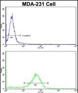
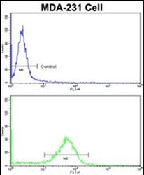
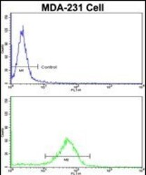
- Flow cytometry analysis of BARON in MDA-231 cells (bottom histogram) compared to a negative control cell (top histogram). Samples were incubated with BARON polyclonal antibody (Product # PA5-72054) followed by a FITC conjugated goat anti-rabbit secondary antibody.
- Submitted by
- Invitrogen Antibodies (provider)
- Main image
- Experimental details
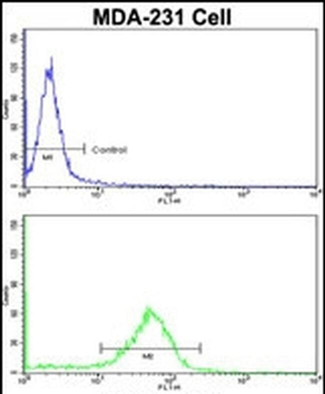
- Flow cytometry of Rubicon in MDA-231 cells (bottom histogram). Samples were incubated with Rubicon polyclonal antibody (Product # PA5-72054) followed by FITC-conjugated goat-anti-rabbit secondary antibody. Negative control cell (top histogram).