 Explore
 Validate
 Learn
Learn Western blot
 Immunohistochemistry
Antibody data
- Antibody Data
- Antigen structure
- References [1]
- Comments [0]
- Validations
- Immunohistochemistry [1]
- Flow cytometry [2]
- Other assay [1]
Submit
Validation data
Reference
Comment
Report error
- Product number
- PA5-23585 - Provider product page

- Provider
- Invitrogen Antibodies
- Product name
- LAPTM5 Polyclonal Antibody
- Antibody type
- Polyclonal
- Antigen
- Synthetic peptide
- Description
- This antibody is predicted to react with bovine based on sequence homology.
- Reactivity
- Human
- Host
- Rabbit
- Isotype
- IgG
- Vial size
- 400 μL
- Concentration
- 2 mg/mL
- Storage
- Store at 4°C short term. For long term storage, store at -20°C, avoiding freeze/thaw cycles.
Submitted references Spine‑specific downregulation of LAPTM5 expression promotes the progression and spinal metastasis of estrogen receptor‑positive breast cancer by activating glutamine‑dependent mTOR signaling.
Meng Q, Zhou L, Liang H, Hu A, Zhou H, Zhou J, Zhou X, Lin H, Li X, Jiang L, Dong J
International journal of oncology 2022 Apr;60(4)
International journal of oncology 2022 Apr;60(4)
No comments: Submit comment
Supportive validation
- Submitted by
- Invitrogen Antibodies (provider)
- Main image
- Experimental details
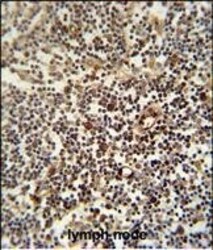
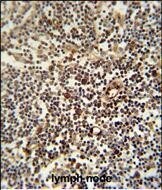
- Immunohistochemistry analysis of LAPTM5 in formalin fixed and paraffin embedded human lymph node. Samples were incubated with LAPTM5 polyclonal antibody (Product # PA5-23585) followed by peroxidase conjugation of the secondary antibody and DAB staining. This data demonstrates the use of this antibody for immunohistochemistry. Clinical relevance has not been evaluated.
Supportive validation
- Submitted by
- Invitrogen Antibodies (provider)
- Main image
- Experimental details
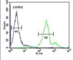
- Flow cytometry analysis of NCI-H460 cells using a LAPTM5 polyclonal antibody (Product # PA5-23585) (right) compared to a negative control cell (left) at a dilution of 1:10-50, followed by a FITC-conjugated goat anti-rabbit antibody

- Submitted by
- Invitrogen Antibodies (provider)
- Main image
- Experimental details
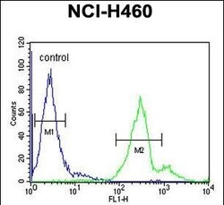
- Flow cytometry of LAPTM5 in NCI-H460 cells (right histogram). Samples were incubated with LAPTM5 polyclonal antibody (Product # PA5-23585) followed by FITC-conjugated goat-anti-rabbit secondary antibody. Negative control cell (left histogram).

Supportive validation
- Submitted by
- Invitrogen Antibodies (provider)
- Main image
- Experimental details
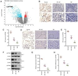
- Figure 1 LAPTM5 expression is downregulated in ER + BC, especially in SM tissue. (A) A volcano plot of the data (GSE14661) was obtained from GEO datasets. Each point represents a gene. Data points highlighted in blue represent genes that are downregulated in SM tissue. The volcano plot shows that LAPTM5 expression is significantly downregulated in SM tissue compared with that in primary ER + BC tissue. (B) IHC staining of LAPTM5 in human ER + BC tissue and its tumor-adjacent normal tissue and SM tissue. (C) Quantitative analysis of the IHC results of LAPTM5 in B. (D) IHC staining of LAPTM5 in mouse ER + BC tissue, tumor-adjacent normal tissue and SM tissue. (E) Quantitative analysis of the IHC results of LAPTM5 in D. (F and G) Western blot and quantitative analysis of LAPTM5 in human ER + BC tissue, tumor-adjacent normal tissue and SM tissue. (H) qPCR results of laptm5 in mouse ER + BC and SM tissue. ** P
