 Explore
 Validate
 Learn
Learn Western blot
Antibody data
- Antibody Data
- Antigen structure
- References [0]
- Comments [0]
- Validations
- Western blot [2]
- Immunocytochemistry [1]
Submit
Validation data
Reference
Comment
Report error
- Product number
- MA5-24275 - Provider product page

- Provider
- Invitrogen Antibodies
- Product name
- ONECUT2 Monoclonal Antibody (606014)
- Antibody type
- Monoclonal
- Antigen
- Recombinant full-length protein
- Description
- No cross-reactivity with recombinant human ONECUT is observed. Reconstitute in sterile PBS to a final concentration of 0.5 mg/mL.
- Reactivity
- Human
- Host
- Mouse
- Isotype
- IgG
- Antibody clone number
- 606014
- Vial size
- 100 µg
- Concentration
- 0.5 mg/mL
- Storage
- -20° C, Avoid Freeze/Thaw Cycles
No comments: Submit comment
Supportive validation
- Submitted by
- Invitrogen Antibodies (provider)
- Main image
- Experimental details
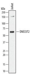
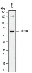
- Western blot analysis from lysates of Jurkat human acute T cell leukemia cell line. PVDF Membrane was probed with 2 µg/mL of mouse Anti-human ONECUT2 Monoclonal Antibody (Product # MA5-24275) followed by HRP-conjugated Anti-mouse IgG Secondary Antibody. A specific band was detected for ONECUT2 at approximately 60 kDa (as indicated). This experiment was conducted under reducing conditions.
- Submitted by
- Invitrogen Antibodies (provider)
- Main image
- Experimental details
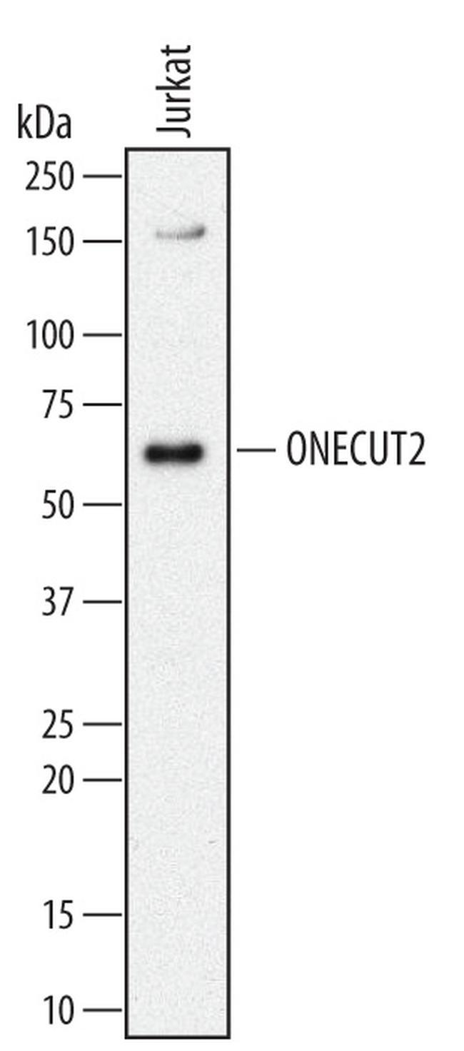
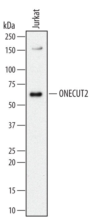
- Western blot analysis of ONECUT2 in Jurkat human acute T cell leukemia cell line. Samples were incubated in ONECUT2 monoclonal antibody (Product # MA5-24275) using a dilution of 2 µg/mL followed by a HRP-conjugated Anti-Mouse IgG secondary antibody. A specific band was detected for PLA2G2A at approximately 17 kDa (as indicated). This experiment was conducted under non-reducing conditions.
Supportive validation
- Submitted by
- Invitrogen Antibodies (provider)
- Main image
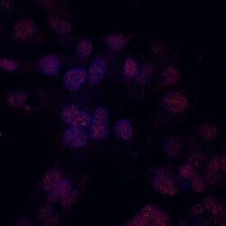
- Experimental details
- Immunocytochemistry analysis of ONECUT2 in immersion fixed HepG2 human hepatocellular carcinoma cell line. Samples were incubated in ONECUT2 monoclonal antibody (Product # MA5-24275) using a dilution of 10 µg/mL for 3 hours at room temperature followed by NorthernLights™ 557-conjugated Anti-Mouse IgG Secondary Antibody (red) and counterstained with DAPI (blue). Specific staining was localized to nuclei.
