Explore
 Validate
 Learn
Learn Western blot
Antibody data
- Antibody Data
- Antigen structure
- References [0]
- Comments [0]
- Validations
- Western blot [3]
- Immunohistochemistry [3]
Submit
Validation data
Reference
Comment
Report error
- Product number
- STJ93515 - Provider product page

- Provider
- St John's Laboratory
- Product name
- Anti-Histone H1 antibody
- Antibody type
- Polyclonal
- Reactivity
- Human, Mouse
- Host
- Rabbit
- Conjugate
- Unconjugated
- Epitope
- 1-80 aa
- Isotype
- IgG
- Vial size
- 200μl,50μl,100μl
- Concentration
- 1 mg/ml
- Storage
- Store at -20°C, and avoid repeat freeze-thaw cycles.
No comments: Submit comment
Supportive validation
Supportive validation
Supportive validation
- Submitted by
- St John's Laboratory (provider)
- Main image
- Experimental details
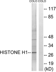
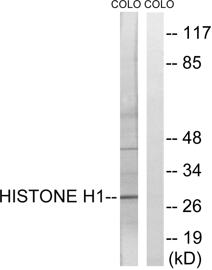
- Western blot analysis of lysates from COLO cells, using Histone H1 Antibody. The lane on the right is blocked with the synthesized peptide.
- Sample type
- NA
- Validation comment
- NA
- Primary Ab dilution
- NA
- Other comments
- NA
- Secondary Ab
- NA
- Secondary Ab dilution
- NA
- Protocol
- NA
Supportive validation
- Submitted by
- St John's Laboratory (provider)
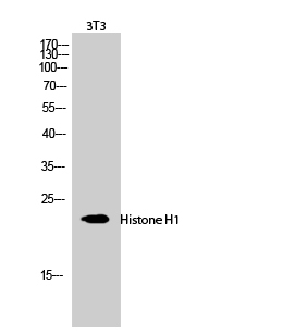
- Main image
- Experimental details
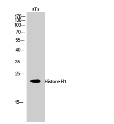
- Western blot analysis of 3T3 cells using Histone H1 Polyclonal Antibody diluted at 1ï¼1000 cells nucleus extracted by Minute TM Cytoplasmic and Nuclear Fractionation kit (SC-003, Inventbiotech, MN, USA).
- Sample type
- NA
- Validation comment
- NA
- Primary Ab dilution
- NA
- Other comments
- NA
- Secondary Ab
- NA
- Secondary Ab dilution
- NA
- Protocol
- NA
Supportive validation
- Submitted by
- St John's Laboratory (provider)
- Main image
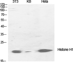
- Experimental details
- Western blot analysis of various cells using Histone H1 Polyclonal Antibody diluted at 1ï¼1000 cells nucleus extracted by Minute TM Cytoplasmic and Nuclear Fractionation kit (SC-003, Inventbiotech, MN, USA).
- Sample type
- NA
- Validation comment
- NA
- Primary Ab dilution
- NA
- Other comments
- NA
- Secondary Ab
- NA
- Secondary Ab dilution
- NA
- Protocol
- NA

Supportive validation
Supportive validation
Supportive validation
- Submitted by
- St John's Laboratory (provider)
- Main image
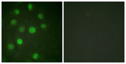
- Experimental details
- Immunofluorescence analysis of HUVEC cells, using Histone H1 Antibody. The picture on the right is blocked with the synthesized peptide.
- Sample type
- NA
- Validation comment
- NA
- Primary Ab dilution
- NA
- Other comments
- NA
- Secondary Ab
- NA
- Secondary Ab dilution
- NA
- Protocol
- NA
Supportive validation
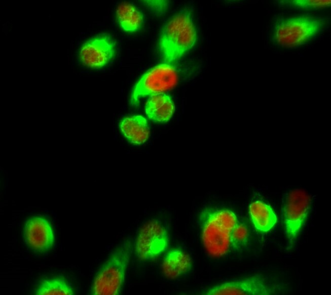
- Submitted by
- St John's Laboratory (provider)
- Main image

- Experimental details
- Immunofluorescence analysis of Hela cell. 1, Histone H1 Polyclonal Antibody (red) was diluted at 1:200 (4°C overnight). Alpha-SMA monoclonal antibody (6A12) (green) was diluted at 1:200 (4°C overnight). 2, Goat Anti Rabbit Alexa Fluor 594 Catalog: (NA was diluted at 1:1000 (room temperature, 50min). Goat Anti Mouse Alexa Fluor 488 Catalog: (NA was diluted at 1:1000 (room temperature, 50min).
- Sample type
- NA
- Validation comment
- NA
- Primary Ab dilution
- NA
- Other comments
- NA
- Secondary Ab
- NA
- Secondary Ab dilution
- NA
- Protocol
- NA
Supportive validation
- Submitted by
- St John's Laboratory (provider)
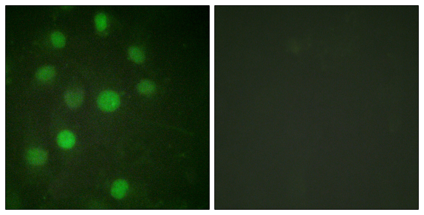
- Main image

- Experimental details
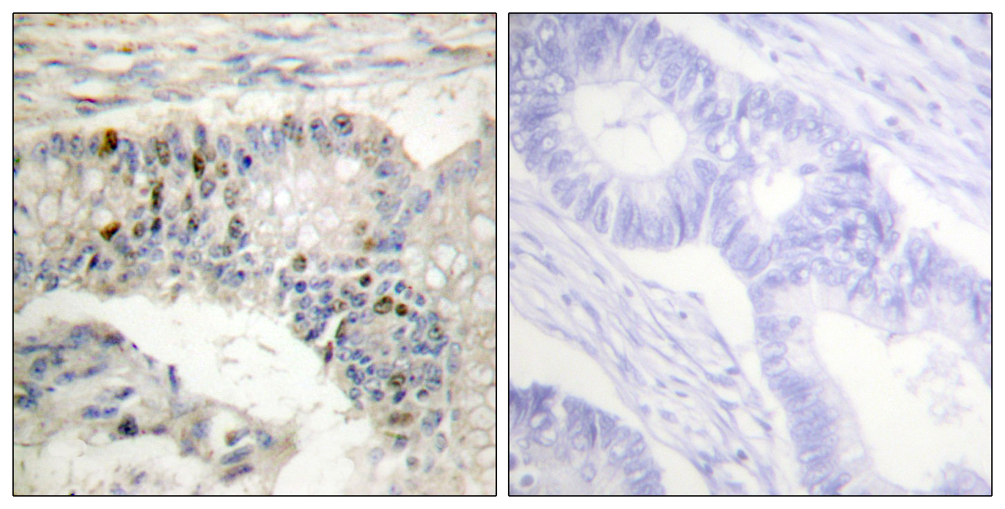
- Immunohistochemistry analysis of paraffin-embedded human colon carcinoma tissue, using Histone H1 Antibody. The picture on the right is blocked with the synthesized peptide.
- Sample type
- NA
- Validation comment
- NA
- Primary Ab dilution
- NA
- Other comments
- NA
- Secondary Ab
- NA
- Secondary Ab dilution
- NA
- Protocol
- NA