 Explore
 Validate
 Learn
Learn Immunocytochemistry
Antibody data
- Antibody Data
- Antigen structure
- References [3]
- Comments [0]
- Validations
- Immunocytochemistry [1]
- Flow cytometry [1]
Submit
Validation data
Reference
Comment
Report error
- Product number
- MAB4617 - Provider product page

- Provider
- R&D Systems
- Product name
- Human GPR34 Antibody
- Antibody type
- Monoclonal
- Description
- Protein A or G purified from hybridoma culture supernatant. Stains human GPR34 transfectants but not irrelevant transfectants.
- Reactivity
- Human
- Host
- Mouse
- Conjugate
- Unconjugated
- Antigen sequence
Q9UPC5- Isotype
- IgG
- Antibody clone number
- 419859
- Vial size
- 100 ug
- Concentration
- LYOPH
- Storage
- Use a manual defrost freezer and avoid repeated freeze-thaw cycles. 12 months from date of receipt, -20 to -70 °C as supplied. 1 month, 2 to 8 °C under sterile conditions after reconstitution. 6 months, -20 to -70 °C under sterile conditions after reconstitution.
Submitted references Single amino acid substitution in LC-CDR1 induces Russell body phenotype that attenuates cellular protein synthesis through eIF2α phosphorylation and thereby downregulates IgG secretion despite operational secretory pathway traffic.
Topogenesis and cell surface trafficking of GPR34 are facilitated by positive-inside rule that effects through a tri-basic motif in the first intracellular loop.
t(X;14)(p11;q32) in MALT lymphoma involving GPR34 reveals a role for GPR34 in tumor cell growth.
Hasegawa H, Hsu A, Tinberg CE, Siegler KE, Nazarian AA, Tsai MM
mAbs 2017 Jul;9(5):854-873
mAbs 2017 Jul;9(5):854-873
Topogenesis and cell surface trafficking of GPR34 are facilitated by positive-inside rule that effects through a tri-basic motif in the first intracellular loop.
Hasegawa H, Patel N, Ettehadieh E, Li P, Lim AC
Biochimica et biophysica acta 2016 Jul;1863(7 Pt A):1534-51
Biochimica et biophysica acta 2016 Jul;1863(7 Pt A):1534-51
t(X;14)(p11;q32) in MALT lymphoma involving GPR34 reveals a role for GPR34 in tumor cell growth.
Ansell SM, Akasaka T, McPhail E, Manske M, Braggio E, Price-Troska T, Ziesmer S, Secreto F, Fonseca R, Gupta M, Law M, Witzig TE, Dyer MJ, Dogan A, Cerhan JR, Novak AJ
Blood 2012 Nov 8;120(19):3949-57
Blood 2012 Nov 8;120(19):3949-57
No comments: Submit comment
Supportive validation
- Submitted by
- R&D Systems (provider)
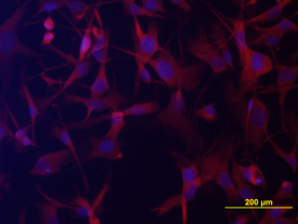
- Main image

- Experimental details
- GPR34 in A172 Human Cell Line. GPR34 was detected in immersion fixed A172 human glioblastoma cell line using Mouse Anti-Human GPR34 Monoclonal Antibody (Catalog # MAB4617) at 10 µg/mL for 3 hours at room temperature. Cells were stained using the NorthernLights™ 557-conjugated Anti-Mouse IgG Secondary Antibody (red; Catalog # NL007) and counterstained with DAPI (blue). View our protocol for Fluorescent ICC Staining of Cells on Coverslips.
Supportive validation
- Submitted by
- R&D Systems (provider)
- Main image

- Experimental details
- Detection of GPR34 in A172 Human Cell Line by Flow Cytometry. A172 human glioblastoma cell line was stained with Mouse Anti-Human GPR34 Monoclonal Antibody (Catalog # MAB4617, filled histogram) or isotype control antibody (Catalog # MAB003, open histogram), followed by Phycoerythrin-conjugated Anti-Mouse IgG F(ab')2 Secondary Antibody (Catalog # F0102B). To facilitate intracellular staining, cells were fixed with paraformaldehyde and permeabilized with saponin.
