 Explore
 Validate
 Learn
Learn Immunocytochemistry
 Immunohistochemistry
Antibody data
- Antibody Data
- Antigen structure
- References [0]
- Comments [0]
- Validations
- Immunocytochemistry [3]
Submit
Validation data
Reference
Comment
Report error
- Product number
- GTX80798 - Provider product page

- Provider
- GeneTex
- Proper citation
- GeneTex Cat#GTX80798, RRID:AB_625832
- Product name
- VR1/ TRPV1 antibody
- Antibody type
- Polyclonal
- Reactivity
- Human, Mouse, Rat
- Host
- Guinea Pig
No comments: Submit comment
Supportive validation
- Submitted by
- GeneTex (provider)
- Main image

- Experimental details
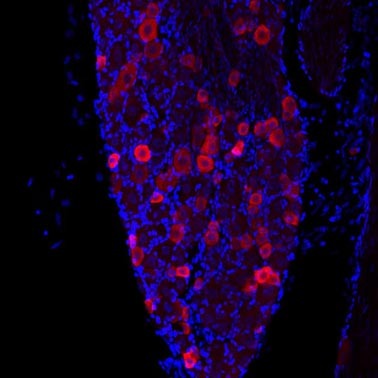
- TRPV1 staining of mouse inferior olive using Cy3-conjugated donkey anti-rabbit secondary antibodies (red color) and DAPI (blue) as a nuclear counterstain.
- Submitted by
- GeneTex (provider)
- Main image

- Experimental details
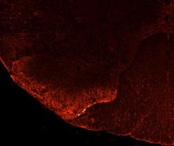
- VR1-C staining of rat dorsal horn (dilution 1:100).
- Submitted by
- GeneTex (provider)
- Main image

- Experimental details
- Evaluation of the expression patterns of TLR4 and CD14 in trigeminal sensory neurons. White arrows depict examples of neurons expressing both markers for each row of three images, and yellow arrows depict examples of neurons that express one but not both markers. Human trigeminal neurons were evaluated for co-localization of TLR4 (Panels A,D), CD14 (Panel B), with a marker for the capsaicinsensitive subclass of nociceptors (TRPV1, Panels B,C for TLR4 and Panels K,L for CD14), or a marker of myelinated sensory neurons (N52, Panels Figure 1. Evaluation of the expression patterns of TLR4 and CD14 in trigeminal sensory neurons. White arrows depict examples of neurons expressing both markers for each row of three images, and yellow arrows depict examples of neurons that express one but not both markers. Human trigeminal neurons were evaluated for co-localization of TLR4 (Panels A,D), CD14 (Panel J), with a marker for the capsaicinsensitive subclass of nociceptors (TRPV1, Panels B,C for TLR4 and Panels K,L for CD14), or a marker of myelinated sensory neurons (N52, Panels E,F). Rat trigeminal neurons were evaluated for co-localization of TLR4 with TRPV1 (Panels G-I) and CD14 with TRPV1 (Panels M-O). The addition of blocking peptide or the deletion of primary or secondary antisera produced a complete loss of signal in both the human and rat tissues. Scale bar is 100 um for Panels A-F and J-L, and 200 um for Panels G-I and M-O. Rat trigeminal neurons were evaluated for co-localization of TLR4 with TRPV1 (Panels G-I) and CD14 with TRPV1 (Panels M-O). The addition of blocking peptide or the deletion of primary or secondary antisera produced a complete loss of signal in both the human and rat tissues. Scale bar is 100 um for Panels A-F and J-L, and 200 um for Panels G-I and M-O
