Explore
 Validate
 Learn
Learn Western blot
 Immunoprecipitation
 Immunohistochemistry
Antibody data
- Antibody Data
- Antigen structure
- References [4]
- Comments [0]
- Validations
- Immunohistochemistry [2]
- Other assay [4]
Submit
Validation data
Reference
Comment
Report error
- Product number
- PA5-29975 - Provider product page

- Provider
- Invitrogen Antibodies
- Product name
- ATP Synthase gamma Polyclonal Antibody
- Antibody type
- Polyclonal
- Antigen
- Recombinant full-length protein
- Description
- Recommended positive controls: HeLa, mouse brain, rat kidney. Predicted reactivity: Mouse (89%), Rat (87%), Bovine (86%). Store product as a concentrated solution. Centrifuge briefly prior to opening the vial.
- Reactivity
- Human, Mouse, Rat, Zebrafish
- Host
- Rabbit
- Isotype
- IgG
- Vial size
- 100 μL
- Concentration
- 0.44 mg/mL
- Storage
- Store at 4°C short term. For long term storage, store at -20°C, avoiding freeze/thaw cycles.
Submitted references The mitochondrial chaperone TRAP1 regulates F-ATP synthase channel formation.
The Unique Cysteine of F-ATP Synthase OSCP Subunit Participates in Modulation of the Permeability Transition Pore.
Toxoplasma gondii GRA8-derived peptide immunotherapy improves tumor targeting of colorectal cancer.
Toxoplasma gondii GRA8 induces ATP5A1-SIRT3-mediated mitochondrial metabolic resuscitation: a potential therapy for sepsis.
Cannino G, Urbani A, Gaspari M, Varano M, Negro A, Filippi A, Ciscato F, Masgras I, Gerle C, Tibaldi E, Brunati AM, Colombo G, Lippe G, Bernardi P, Rasola A
Cell death and differentiation 2022 Dec;29(12):2335-2346
Cell death and differentiation 2022 Dec;29(12):2335-2346
The Unique Cysteine of F-ATP Synthase OSCP Subunit Participates in Modulation of the Permeability Transition Pore.
Carraro M, Jones K, Sartori G, Schiavone M, Antonucci S, Kucharczyk R, di Rago JP, Franchin C, Arrigoni G, Forte M, Bernardi P
Cell reports 2020 Sep 1;32(9):108095
Cell reports 2020 Sep 1;32(9):108095
Toxoplasma gondii GRA8-derived peptide immunotherapy improves tumor targeting of colorectal cancer.
Kim JS, Lee D, Kim D, Mun SJ, Cho E, Son W, Yang CS
Oncotarget 2020 Jan 7;11(1):62-73
Oncotarget 2020 Jan 7;11(1):62-73
Toxoplasma gondii GRA8 induces ATP5A1-SIRT3-mediated mitochondrial metabolic resuscitation: a potential therapy for sepsis.
Kim YR, Kim JS, Yun JS, Kim S, Kim SY, Jang K, Yang CS
Experimental & molecular medicine 2018 Mar 30;50(3):e464
Experimental & molecular medicine 2018 Mar 30;50(3):e464
No comments: Submit comment
Supportive validation
- Submitted by
- Invitrogen Antibodies (provider)
- Main image
- Experimental details
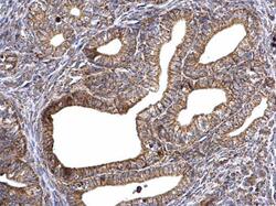
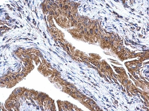
- ATP Synthase gamma Polyclonal Antibody detects ATP synthase gamma protein at mitochondria on mouse cervix by immunohistochemical analysis. Sample: Paraffin-embedded mouse cervix. ATP Synthase gamma Polyclonal Antibody (Product # PA5-29975) dilution: 1:500. Antigen Retrieval: EDTA based buffer, pH 8.0, 15 min.

- Submitted by
- Invitrogen Antibodies (provider)
- Main image

- Experimental details
- ATP Synthase gamma Polyclonal Antibody detects ATP synthase gamma protein at mitochondria on mouse cervix by immunohistochemical analysis. Sample: Paraffin-embedded mouse cervix. ATP Synthase gamma Polyclonal Antibody (Product # PA5-29975) dilution: 1:500. Antigen Retrieval: EDTA based buffer, pH 8.0, 15 min.
Supportive validation
- Submitted by
- Invitrogen Antibodies (provider)
- Main image

- Experimental details
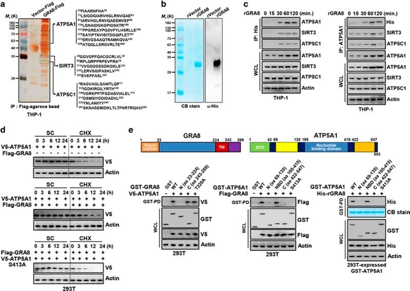
- Figure 1 GRA8 interacts with ATP5A1 and SIRT3. ( a ) Identification of ATP5A1, SIRT3 and ATP5C1 by mass spectrometry analysis in THP-1 cell lysates expressed with GRA8 or vector. ( b ) Bacterially purified 6xHis-GRA8 were analyzed by Coomassie blue staining (left) or immunoblotting (IB) with alphaHis (right). ( c ) THP-1 cells were stimulated with rGRA8 (5 mug ml -1 ) for the indicated times, followed by immunoprecipitation (IP) with alphaHis-agarose bead or alphaATP5A1 and IB with alphaATP5A1, alphaSIRT3, alphaATP5C1, alphaHis and alphaActin. ( d ) GRA8-mediated increases of ATP5A1 stability. At 24 h after transfection with V5-ATP5A1and/or Flag-GRA8, 293T cells were treated with solvent control (SC) or cyclohexamide (CHX, 1 mug ml -1 ) for indicated times and cell lysates were used for IB with alphaV5 and alphaActin. ( e ) Binding mapping. Schematic diagrams of the structures of GRA8 and ATP5A1 (upper). At 48 h after transfection with mammalian glutathione S -transferase (GST) or GST-GRA8 and truncated mutant constructs together with V5-ATP5A1, or GST-ATP5A1 constructs together with Flag or His-GRA8, 293T cells were used for GST pull down, followed by IB with alphaV5, alphaFlag or alphaHis. Whole cell lysates (WCLs) were used for IB with alphaGST, alphaV5, alphaFlag, alphaHis or alphaActin. The data are representative of four independent experiments with similar results ( a - e ).
- Submitted by
- Invitrogen Antibodies (provider)
- Main image
- Experimental details
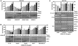
- Figure 1 The rGRA8 treatment increases the HCT116 cell death by mitochondria pathways. ( A and B ) The various cancer cells were incubated with WT rGRA8 (20 mug/mL) and its mutants for the indicated times and then cell viability measured with MTT assay (upper) and subjected to IB with alphaATP5A1, alphaSIRT3, or alphaActin (lower). ( C ) HCT116 cells were transduced with lentivirus-shRNA-NS or lentivirus-shRNA-PKCalpha or SIRT3 (MOI = 100) with polybrene (8 mug/mL) (right) for 2 days. The cells were incubated with WT rGRA8 for the indicated times and subjected to IP with alphaLys-AC or alphaHis and IB with alphaATP5A1, alphaATP5C1, alphaSIRT3, alphaPGC-1, alphaPKCalpha, or alphaActin. The data are representative of five independent experiments with similar results. Statistical significance was determined by Student's t -test with Bonferroni adjustment; ** P < 0.01, *** P < 0.001 compared with rVector-treated.

- Submitted by
- Invitrogen Antibodies (provider)
- Main image

- Experimental details
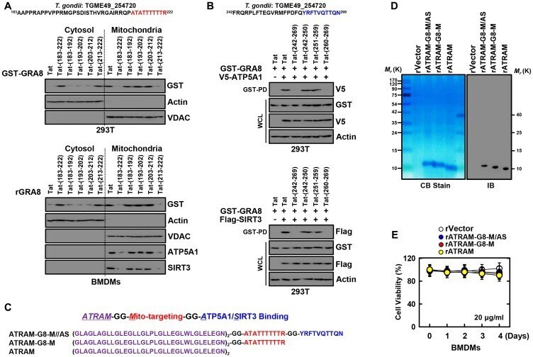
- Figure 2 Design and expression of ATRAM-conjugated multifunctional GRA8 peptide-based protein. ( A ) Subcellular fractionation of 293T cells are expressing GST-GRA8 WT or incubation with WT rGRA8 (1 mug/mL) BMDMs and treated with several Tat-GRA8 peptide (10 µM) for 6 h. Mitochondrial and cytosolic fractions were fractionated and analyzed for expression of GST by IB. Purity of the fractions was assessed by blotting for VDAC (mitochondrial protein) and Actin (cytosolic protein). ( B ) At 12 hr post-transfection with mammalian GST-GRA8 constructs together with V5-ATP5A1, or Flag-SIRT3 and 293T cells treated with several Tat-GRA8 peptide (10 µM) for 6 h. 293T cells were used for GST pulldown, followed by IB with alphaV5 or alphaFlag. WCLs were used for IB with alphaGST, alphaV5, alphaFlag or alphaActin. ( C ) Schematic in design of ATRAM-GRA8-M/AS and its mutants. ( D ) Bacterially purified 6xHis-rATRAM-GRA8-M/AS and its mutants were analyzed by Coomassie blue staining (left) or immuno blotting (IB) with alphaHis (right). ( E ) BMDMs were incubated with rATRAM-GRA8-M/AS and its mutants (20 mug/mL) for the indicated times and then cell viability measured with MTT assay. The data are representative of four independent experiments with similar results (A, B, D, and E).
- Submitted by
- Invitrogen Antibodies (provider)
- Main image

- Experimental details
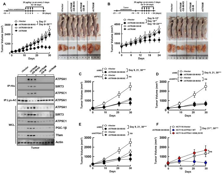
- Figure 4 The rATRAM-GRA8-M/AS showed an antitumor activity in HCT116 xenografts. ( A and B ) Schematic of the xenograft model treated with or rATRAM-GRA8-M/AS or its mutants (upper). HCT116 cells were subcutaneously injected into the flanks of nude mice. The length and width of the tumors were measured using calipers, and the tumor volume was calculated every third day for 30 days. At day 15 after administration, the mice were euthanatized, and the interaction with GRA8, levels of acetylation of ATP5A1, or expression of OXPHOS were analyzed (A, lower). Representative images of tumors from mice treated with rATRAM-GRA8-M/AS or mutants on day 15 (right). ( C-F ) HCT116 cells were transduced with lentivirus-shRNA-NS or lentivirus-shRNA-PKCalpha, SIRT3, or ATP5A1 with polybrene (C-E) or HCT116-expressed ATP5A1 WT and mutant cells were subcutaneously injected into the flanks of nude mice. Individual tumor volumes from each mouse from each group were averaged and plotted against the number days postinoculation. Statistical significance was determined by two-way analysis of variance (ANOVA) with Tukey's posttest; * P < 0.05, ** P < 0.01, *** P < 0.001 compared with rVector. Each group contained ten mice. The data are representative of two independent experiments with similar results.