Explore
 Validate
 Learn
Learn Western blot
 Immunocytochemistry
Antibody data
- Antibody Data
- Antigen structure
- References [1]
- Comments [0]
- Validations
- Immunocytochemistry [1]
Submit
Validation data
Reference
Comment
Report error
- Product number
- AMAb91394 - Provider product page

- Provider
- Atlas Antibodies
- Proper citation
- Atlas Antibodies Cat#AMAb91394, RRID:AB_2716670
- Product name
- Anti-TUBB3
- Antibody type
- Monoclonal
- Description
- Monoclonal Antibody against Human TUBB3, Clone ID: CL5813, Gene description: Tubulin beta 3 class iii, Alternative Gene Names: beta-4, CFEOM3, CFEOM3A, FEOM3, Validated applications: ICC, WB, IHC, Uniprot ID: Q13509, Storage: Store at +4°C for short term storage. Long time storage is recommended at -20°C.
- Reactivity
- Human, Mouse, Rat
- Host
- Mouse
- Conjugate
- Unconjugated
- Isotype
- IgG
- Antibody clone number
- CL5813
- Vial size
- 100 µl
- Concentration
- 1.0 mg/ml
- Storage
- Store at +4°C for short term storage. Long time storage is recommended at -20°C.
- Handling
- The antibody solution should be gently mixed before use.
Submitted references Laminar dynamics of deep projection neurons and mode of subplate formation are hallmarks of histogenetic subdivisions of the human cingulate cortex before onset of arealization
Junaković A, Kopić J, Duque A, Rakic P, Krsnik Ž, Kostović I
Brain Structure and Function 2023;228(2):613-633
Brain Structure and Function 2023;228(2):613-633
No comments: Submit comment
Supportive validation
- Submitted by
- Atlas Antibodies (provider)
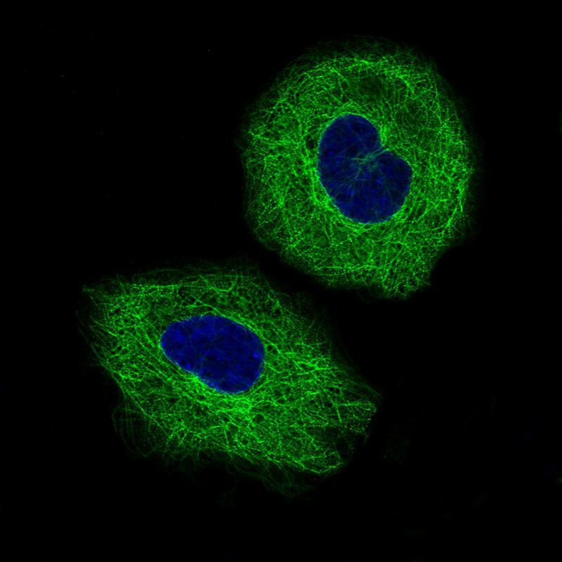
- Main image

- Experimental details
- Immunofluorescence staining of A549 cells using the Anti-TUBB3 monoclonal antibody, showing specific staining on microtubules in green. Microtubule- and nuclear probes are visualized in red and blue, respectively (where available).
- Sample type
- Human