 Explore
 Validate
 Learn
Learn Western blot
Antibody data
- Antibody Data
- Antigen structure
- References [0]
- Comments [0]
- Validations
- Western blot [1]
- Immunohistochemistry [3]
Submit
Validation data
Reference
Comment
Report error
- Product number
- GTX133149 - Provider product page

- Provider
- GeneTex
- Product name
- IP3 Receptor I antibody
- Antibody type
- Polyclonal
- Reactivity
- Mouse, Rat
- Host
- Rabbit
No comments: Submit comment
Supportive validation
- Submitted by
- GeneTex (provider)
- Main image
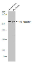
- Experimental details
- Various tissue extracts (50 ?g) were separated by 5% SDS-PAGE, and the membrane was blotted with IP3 Receptor I antibody (GTX133149) diluted at 1:500. The signal was developed with Trident ECL plus-Enhanced.

Supportive validation
- Submitted by
- GeneTex (provider)
- Main image

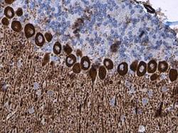
- Experimental details
- IP3 Receptor I antibody detects IP3 Receptor I protein at cytoplasm in mouse brain by immunohistochemical analysis. Sample: Paraffin-embedded mouse brain. IP3 Receptor I antibody (GTX133149) diluted at 1:500.

- Submitted by
- GeneTex (provider)
- Main image
- Experimental details
- IP3 Receptor I antibody detects IP3 Receptor I protein at cytoplasm in mouse brain by immunohistochemical analysis. Sample: Paraffin-embedded mouse brain. IP3 Receptor I antibody (GTX133149) diluted at 1:500.

- Submitted by
- GeneTex (provider)
- Main image

- Experimental details
- IP3 Receptor I antibody detects IP3 Receptor I protein expression by immunohistochemical analysis.Sample: Frozen-sectioned adult mouse cerebellum. Green: IP3 Receptor I protein stained by IP3 Receptor I antibody (GTX133149) diluted at 1:250.Red: beta Tubulin 3/ TUJ1, stained by beta Tubulin 3/ TUJ1 antibody [GT11710] (GTX631836) diluted at 1:500.Blue: Fluoroshield with DAPI (GTX30920).
