Explore
 Validate
 Learn
Learn Immunohistochemistry
Antibody data
- Antibody Data
- Antigen structure
- References [48]
- Comments [0]
- Validations
- Immunohistochemistry [1]
Submit
Validation data
Reference
Comment
Report error
- Product number
- HPA051870 - Provider product page

- Provider
- Atlas Antibodies
- Proper citation
- Atlas Antibodies Cat#HPA051870, RRID:AB_2681645
- Product name
- Anti-TMEM119
- Antibody type
- Polyclonal
- Description
- Polyclonal Antibody against Human TMEM119, Gene description: transmembrane protein 119, Validated applications: IHC, Uniprot ID: Q4V9L6, Storage: Store at +4°C for short term storage. Long time storage is recommended at -20°C.
- Reactivity
- Human
- Host
- Rabbit
- Conjugate
- Unconjugated
- Isotype
- IgG
- Vial size
- 100 µl
- Concentration
- 0.1 mg/ml
- Storage
- Store at +4°C for short term storage. Long time storage is recommended at -20°C.
- Handling
- The antibody solution should be gently mixed before use.
Submitted references Human Microglia–Like Cells Differentiated from Monocytes with GM-CSF and IL-34 Show Phagocytosis of α-Synuclein Aggregates and C/EBPβ-Dependent Proinflammatory Activation
A glia-enriched stem cell 3D model of the human brain mimics the glial-immune neurodegenerative phenotypes of multiple sclerosis
Myeloid cell-derived creatine in the hypoxic niche promotes glioblastoma growth
Bone marrow-derived myeloid cells transiently colonize the brain during postnatal development and interact with glutamatergic synapses
CHIT1 at diagnosis predicts faster disability progression and reflects early microglial activation in multiple sclerosis
Ultrasound-mediated delivery of doxorubicin to the brain results in immune modulation and improved responses to PD-1 blockade in gliomas
Landscape of brain myeloid cell transcriptome along the spatiotemporal progression of Alzheimer’s disease reveals distinct sequential responses to Aβ and tau
The CXCL16-CXCR6 axis in glioblastoma modulates T-cell activity in a spatiotemporal context
Human-induced pluripotent stem cell-derived microglia integrate into mouse retina and recapitulate features of endogenous microglia
ATP1A3 as a target for isolating neuron-specific extracellular vesicles from human brain and biofluids
A potential protective role of the nuclear receptor-related factor 1 (Nurr1) in multiple sclerosis motor cortex: a neuropathological study
Tau activation of microglial cGAS–IFN reduces MEF2C-mediated cognitive resilience
GBMdeconvoluteR accurately infers proportions of neoplastic and immune cell populations from bulk glioblastoma transcriptomics data
Cerebellar presence of immune cells in patients with neuro-coeliac disease
The localization of molecularly distinct microglia populations to Alzheimer's disease pathologies using QUIVER
A systematic characterization of microglia-like cell occurrence during retinal organoid differentiation
Immunopathology of the optic nerve in multiple sclerosis
Dominant-acting CSF1R variants cause microglial depletion and altered astrocytic phenotype in zebrafish and adult-onset leukodystrophy
Advances in Visualizing Microglial Cells in Human Central Nervous System Tissue
Molecular Signature of Neuroinflammation Induced in Cytokine-Stimulated Human Cortical Spheroids
Changes in glial cell phenotypes precede overt neurofibrillary tangle formation, correlate with markers of cortical cell damage, and predict cognitive status of individuals at Braak III-IV stages
Apolipoprotein E4 impairs the response of neurodegenerative retinal microglia and prevents neuronal loss in glaucoma.
Activated microglia do not increase 18 kDa translocator protein (TSPO) expression in the multiple sclerosis brain
MicroRNA‐34a activation in tuberous sclerosis complex during early brain development may lead to impaired corticogenesis
The influence of HLA‐DRB1*15 on the relationship between microglia and neurons in multiple sclerosis normal appearing cortical grey matter
Meningeal inflammation in multiple sclerosis induces phenotypic changes in cortical microglia that differentially associate with neurodegeneration
A loss of mature microglial markers without immune activation in schizophrenia.
VISTA regulates microglia homeostasis and myelin phagocytosis, and is associated with MS lesion pathology.
Poly-ADP-ribosylation drives loss of protein homeostasis in ATM and Mre11 deficiency.
Microglia promote glioblastoma via mTOR‐mediated immunosuppression of the tumour microenvironment
Distinct microglial and macrophage distribution patterns in the concentric and lamellar lesions in Baló's disease and neuromyelitis optica spectrum disorders
Increased expression of miR142 and miR155 in glial and immune cells after traumatic brain injury may contribute to neuroinflammation via astrocyte activation
Microglia express TMEM119 in the brains of Nasu-Hakola disease
Multiplexed imaging of immune cells in staged multiple sclerosis lesions by mass cytometry
Microglia damage precedes major myelin breakdown in X‐linked adrenoleukodystrophy and metachromatic leukodystrophy
Single-Cell RNA Sequencing of Microglia throughout the Mouse Lifespan and in the Injured Brain Reveals Complex Cell-State Changes
A quantitative neuropathological assessment of translocator protein expression in multiple sclerosis
Regulation of microglial TMEM119 and P2RY12 immunoreactivity in multiple sclerosis white and grey matter lesions is dependent on their inflammatory environment.
TLR-stimulated IRAKM activates caspase-8 inflammasome in microglia and promotes neuroinflammation
TMEM119 silencing inhibits cell viability and causes the apoptosis of gastric cancer SGC-7901 cells
The TREM2-APOE Pathway Drives the Transcriptional Phenotype of Dysfunctional Microglia in Neurodegenerative Diseases
Dominant role of microglial and macrophage innate immune responses in human ischemic infarcts
Transcriptomic analysis of purified human cortical microglia reveals age-associated changes
Loss of ‘homeostatic’ microglia and patterns of their activation in active multiple sclerosis
Llaves-López A, Micoli E, Belmonte-Mateos C, Aguilar G, Alba C, Marsal A, Pulido-Salgado M, Rabaneda-Lombarte N, Solà C, Serratosa J, Vidal-Taboada J, Saura J
Molecular Neurobiology 2024;62(1):756-772
Molecular Neurobiology 2024;62(1):756-772
A glia-enriched stem cell 3D model of the human brain mimics the glial-immune neurodegenerative phenotypes of multiple sclerosis
Fagiani F, Pedrini E, Taverna S, Brambilla E, Murtaj V, Podini P, Ruffini F, Butti E, Braccia C, Andolfo A, Magliozzi R, Smirnova L, Kuhlmann T, Quattrini A, Calabresi P, Reich D, Martino G, Panina-Bordignon P, Absinta M
Cell Reports Medicine 2024;5(8):101680
Cell Reports Medicine 2024;5(8):101680
Myeloid cell-derived creatine in the hypoxic niche promotes glioblastoma growth
Rashidi A, Billingham L, Zolp A, Chia T, Silvers C, Katz J, Park C, Delay S, Boland L, Geng Y, Markwell S, Dmello C, Arrieta V, Zilinger K, Jacob I, Lopez-Rosas A, Hou D, Castro B, Steffens A, McCortney K, Walshon J, Flowers M, Lin H, Wang H, Zhao J, Sonabend A, Zhang P, Ahmed A, Brat D, Heiland D, Lee-Chang C, Lesniak M, Chandel N, Miska J
Cell Metabolism 2024;36(1):62-77.e8
Cell Metabolism 2024;36(1):62-77.e8
Bone marrow-derived myeloid cells transiently colonize the brain during postnatal development and interact with glutamatergic synapses
Carrier M, Robert M, St-Pierre M, Ibáñez F, Gonçalves de Andrade E, Laroche A, Picard K, Vecchiarelli H, Savage J, Boilard É, Desjardins M, Tremblay M
iScience 2024;27(7):110037
iScience 2024;27(7):110037
CHIT1 at diagnosis predicts faster disability progression and reflects early microglial activation in multiple sclerosis
Beliën J, Swinnen S, D’hondt R, Verdú de Juan L, Dedoncker N, Matthys P, Bauer J, Vens C, Moylett S, Dubois B
Nature Communications 2024;15(1)
Nature Communications 2024;15(1)
Ultrasound-mediated delivery of doxorubicin to the brain results in immune modulation and improved responses to PD-1 blockade in gliomas
Arrieta V, Gould A, Kim K, Habashy K, Dmello C, Vázquez-Cervantes G, Palacín-Aliana I, McManus G, Amidei C, Gomez C, Dhiantravan S, Chen L, Zhang D, Saganty R, Cholak M, Pandey S, McCord M, McCortney K, Castro B, Ward R, Muzzio M, Bouchoux G, Desseaux C, Canney M, Carpentier A, Zhang B, Miska J, Lesniak M, Horbinski C, Lukas R, Stupp R, Lee-Chang C, Sonabend A
Nature Communications 2024;15(1)
Nature Communications 2024;15(1)
Landscape of brain myeloid cell transcriptome along the spatiotemporal progression of Alzheimer’s disease reveals distinct sequential responses to Aβ and tau
Wachter A, Woodbury M, Lombardo S, Abdourahman A, Wuest C, McGlame E, Pastika T, Tamm J, Romanul N, Yanamandra K, Bennett R, Lin G, Kwon T, Liao F, Klein C, Grinberg Y, Jaisa-aad M, Li H, Frosch M, Kummer M, Das S, Dellovade T, Karran E, Langlois X, Ried J, Serrano-Pozo A, Talanian R, Biber K, Hyman B
Acta Neuropathologica 2024;147(1)
Acta Neuropathologica 2024;147(1)
The CXCL16-CXCR6 axis in glioblastoma modulates T-cell activity in a spatiotemporal context
Chia T, Billingham L, Boland L, Katz J, Arrieta V, Shireman J, Rosas A, DeLay S, Zillinger K, Geng Y, Kruger J, Silvers C, Wang H, Vazquez Cervantes G, Hou D, Wang S, Wan H, Sonabend A, Zhang P, Lee-Chang C, Miska J
Frontiers in Immunology 2024;14
Frontiers in Immunology 2024;14
Human-induced pluripotent stem cell-derived microglia integrate into mouse retina and recapitulate features of endogenous microglia
Ma W, Zhao L, Xu B, Fariss R, Redmond T, Zou J, Wong W, Li W
eLife 2024;12
eLife 2024;12
Thompson J, Nelson E, Tippani M, Ramnauth A, Divecha H, Miller R, Eagles N, Pattie E, Kwon S, Bach S, Kaipa U, Yao J, Hou C, Kleinman J, Collado-Torres L, Han S, Maynard K, Hyde T, Martinowich K, Page S, Hicks S
2024
2024
Huuki-Myers L, Montgomery K, Kwon S, Cinquemani S, Eagles N, Gonzalez-Padilla D, Maden S, Kleinman J, Hyde T, Hicks S, Maynard K, Collado-Torres L
2024
2024
ATP1A3 as a target for isolating neuron-specific extracellular vesicles from human brain and biofluids
You Y, Zhang Z, Sultana N, Ericsson M, Martens Y, Sun M, Kanekiyo T, Ikezu S, Shaffer S, Ikezu T
Science Advances 2023;9(37)
Science Advances 2023;9(37)
A potential protective role of the nuclear receptor-related factor 1 (Nurr1) in multiple sclerosis motor cortex: a neuropathological study
Pansieri J, Pisa M, Yates R, Esiri M, DeLuca G
Brain Communications 2023;5(2)
Brain Communications 2023;5(2)
Tau activation of microglial cGAS–IFN reduces MEF2C-mediated cognitive resilience
Udeochu J, Amin S, Huang Y, Fan L, Torres E, Carling G, Liu B, McGurran H, Coronas-Samano G, Kauwe G, Mousa G, Wong M, Ye P, Nagiri R, Lo I, Holtzman J, Corona C, Yarahmady A, Gill M, Raju R, Mok S, Gong S, Luo W, Zhao M, Tracy T, Ratan R, Tsai L, Sinha S, Gan L
Nature Neuroscience 2023;26(5):737-750
Nature Neuroscience 2023;26(5):737-750
GBMdeconvoluteR accurately infers proportions of neoplastic and immune cell populations from bulk glioblastoma transcriptomics data
Ajaib S, Lodha D, Pollock S, Hemmings G, Finetti M, Gusnanto A, Chakrabarty A, Ismail A, Wilson E, Varn F, Hunter B, Filby A, Brockman A, McDonald D, Verhaak R, Ihrie R, Stead L
Neuro-Oncology 2023;25(7):1236-1248
Neuro-Oncology 2023;25(7):1236-1248
Cerebellar presence of immune cells in patients with neuro-coeliac disease
Rouvroye M, Bontkes H, Bol J, Lissenberg-Witte B, Byrnes V, Bennani F, Jordanova E, Wilhelmus M, Mulder C, van der Valk P, Rozemuller A, Bouma G, Van Dam A
Acta Neuropathologica Communications 2023;11(1)
Acta Neuropathologica Communications 2023;11(1)
The localization of molecularly distinct microglia populations to Alzheimer's disease pathologies using QUIVER
Shahidehpour R, Nelson A, Sanders L, Embry C, Nelson P, Bachstetter A
Acta Neuropathologica Communications 2023;11(1)
Acta Neuropathologica Communications 2023;11(1)
Que Z, Olivero-Acosta M, Chen I, Zhang J, Wettschurack K, Wu J, Xiao T, Otterbacher C, Wang M, Harlow H, Cui N, Chen X, Deming B, Halurkar M, Zhao Y, Rochet J, Xu R, Brewster A, Wu L, Yuan C, Skarnes W, Yang Y
2023
2023
A systematic characterization of microglia-like cell occurrence during retinal organoid differentiation
Bartalska K, Hübschmann V, Korkut-Demirbaş M, Cubero R, Venturino A, Rössler K, Czech T, Siegert S
iScience 2022;25(7):104580
iScience 2022;25(7):104580
Immunopathology of the optic nerve in multiple sclerosis
Fernández Blanco L, Marzin M, Leistra A, van der Valk P, Nutma E, Amor S
Clinical and Experimental Immunology 2022;209(2):236-246
Clinical and Experimental Immunology 2022;209(2):236-246
Dominant-acting CSF1R variants cause microglial depletion and altered astrocytic phenotype in zebrafish and adult-onset leukodystrophy
Berdowski W, van der Linde H, Breur M, Oosterhof N, Beerepoot S, Sanderson L, Wijnands L, de Jong P, Tsai-Meu-Chong E, de Valk W, de Witte M, van IJcken W, Demmers J, van der Knaap M, Bugiani M, Wolf N, van Ham T
Acta Neuropathologica 2022;144(2):211-239
Acta Neuropathologica 2022;144(2):211-239
Advances in Visualizing Microglial Cells in Human Central Nervous System Tissue
Uff C, Patel K, Yeung C, Yip P
Biomolecules 2022;12(5):603
Biomolecules 2022;12(5):603
Molecular Signature of Neuroinflammation Induced in Cytokine-Stimulated Human Cortical Spheroids
De Kleijn K, Straasheijm K, Zuure W, Martens G
Biomedicines 2022;10(5):1025
Biomedicines 2022;10(5):1025
Changes in glial cell phenotypes precede overt neurofibrillary tangle formation, correlate with markers of cortical cell damage, and predict cognitive status of individuals at Braak III-IV stages
Taddei R, Sanchez-Mico M, Bonnar O, Connors T, Gaona A, Denbow D, Frosch M, Gómez-Isla T
Acta Neuropathologica Communications 2022;10(1)
Acta Neuropathologica Communications 2022;10(1)
Apolipoprotein E4 impairs the response of neurodegenerative retinal microglia and prevents neuronal loss in glaucoma.
Margeta MA, Yin Z, Madore C, Pitts KM, Letcher SM, Tang J, Jiang S, Gauthier CD, Silveira SR, Schroeder CM, Lad EM, Proia AD, Tanzi RE, Holtzman DM, Krasemann S, Chen DF, Butovsky O
Immunity 2022 Sep 13;55(9):1627-1644.e7
Immunity 2022 Sep 13;55(9):1627-1644.e7
Hou D, Castro B, Dapash M, Zolp A, Katz J, Arrieta V, Biermann J, Melms J, Kueckelhaus J, Benotmane J, Youngblood M, Rashidi A, Billingham L, Dmello C, Vazquez-Cervantes G, Lopez-Rosas A, Han Y, Patel R, Chia T, Sun L, Prins R, Izar B, Heiland D, Zhang P, Sonabend A, Miska J, Lesniak M, Zhao J, Lee-Chang C
2022
2022
Activated microglia do not increase 18 kDa translocator protein (TSPO) expression in the multiple sclerosis brain
Nutma E, Gebro E, Marzin M, van der Valk P, Matthews P, Owen D, Amor S
Glia 2021;69(10):2447-2458
Glia 2021;69(10):2447-2458
MicroRNA‐34a activation in tuberous sclerosis complex during early brain development may lead to impaired corticogenesis
Korotkov A, Sim N, Luinenburg M, Anink J, van Scheppingen J, Zimmer T, Bongaarts A, Broekaart D, Mijnsbergen C, Jansen F, Van Hecke W, Spliet W, van Rijen P, Feucht M, Hainfellner J, Kršek P, Zamecnik J, Crino P, Kotulska K, Lagae L, Jansen A, Kwiatkowski D, Jozwiak S, Curatolo P, Mühlebner A, Lee J, Mills J, van Vliet E, Aronica E
Neuropathology and Applied Neurobiology 2021;47(6):796-811
Neuropathology and Applied Neurobiology 2021;47(6):796-811
The influence of HLA‐DRB1*15 on the relationship between microglia and neurons in multiple sclerosis normal appearing cortical grey matter
Yates R, Pansieri J, Li Q, Bell J, Yee S, Palace J, Esiri M, DeLuca G
Brain Pathology 2021;32(4)
Brain Pathology 2021;32(4)
Meningeal inflammation in multiple sclerosis induces phenotypic changes in cortical microglia that differentially associate with neurodegeneration
van Olst L, Rodriguez-Mogeda C, Picon C, Kiljan S, James R, Kamermans A, van der Pol S, Knoop L, Michailidou I, Drost E, Franssen M, Schenk G, Geurts J, Amor S, Mazarakis N, van Horssen J, de Vries H, Reynolds R, Witte M
Acta Neuropathologica 2021;141(6):881-899
Acta Neuropathologica 2021;141(6):881-899
A loss of mature microglial markers without immune activation in schizophrenia.
Snijders GJLJ, van Zuiden W, Sneeboer MAM, Berdenis van Berlekom A, van der Geest AT, Schnieder T, MacIntyre DJ, Hol EM, Kahn RS, de Witte LD
Glia 2021 May;69(5):1251-1267
Glia 2021 May;69(5):1251-1267
VISTA regulates microglia homeostasis and myelin phagocytosis, and is associated with MS lesion pathology.
Borggrewe M, Kooistra SM, Wesseling EM, Gierschek FL, Brummer ML, Nowak EC, Medeiros-Furquim T, Otto TA, Lee SW, Noelle RJ, Eggen BJL, Laman JD
Acta neuropathologica communications 2021 May 18;9(1):91
Acta neuropathologica communications 2021 May 18;9(1):91
Poly-ADP-ribosylation drives loss of protein homeostasis in ATM and Mre11 deficiency.
Lee JH, Ryu SW, Ender NA, Paull TT
Molecular cell 2021 Apr 1;81(7):1515-1533.e5
Molecular cell 2021 Apr 1;81(7):1515-1533.e5
Microglia promote glioblastoma via mTOR‐mediated immunosuppression of the tumour microenvironment
Dumas A, Pomella N, Rosser G, Guglielmi L, Vinel C, Millner T, Rees J, Aley N, Sheer D, Wei J, Marisetty A, Heimberger A, Bowman R, Brandner S, Joyce J, Marino S
The EMBO Journal 2020;39(15)
The EMBO Journal 2020;39(15)
Distinct microglial and macrophage distribution patterns in the concentric and lamellar lesions in Baló's disease and neuromyelitis optica spectrum disorders
Hayashida S, Masaki K, Suzuki S, Yamasaki R, Watanabe M, Koyama S, Isobe N, Matsushita T, Takahashi K, Tabira T, Iwaki T, Kira J
Brain Pathology 2020;30(6):1144-1157
Brain Pathology 2020;30(6):1144-1157
Increased expression of miR142 and miR155 in glial and immune cells after traumatic brain injury may contribute to neuroinflammation via astrocyte activation
Korotkov A, Puhakka N, Gupta S, Vuokila N, Broekaart D, Anink J, Heiskanen M, Karttunen J, van Scheppingen J, Huitinga I, Mills J, van Vliet E, Pitkänen A, Aronica E
Brain Pathology 2020;30(5):897-912
Brain Pathology 2020;30(5):897-912
Microglia express TMEM119 in the brains of Nasu-Hakola disease
Satoh J, Kino Y, Yanaizu M, Ishida T, Saito Y
Intractable & Rare Diseases Research 2019;8(4):260-265
Intractable & Rare Diseases Research 2019;8(4):260-265
Multiplexed imaging of immune cells in staged multiple sclerosis lesions by mass cytometry
Ramaglia V, Sheikh-Mohamed S, Legg K, Park C, Rojas O, Zandee S, Fu F, Ornatsky O, Swanson E, Pitt D, Prat A, McKee T, Gommerman J
eLife 2019;8
eLife 2019;8
Microglia damage precedes major myelin breakdown in X‐linked adrenoleukodystrophy and metachromatic leukodystrophy
Bergner C, van der Meer F, Winkler A, Wrzos C, Türkmen M, Valizada E, Fitzner D, Hametner S, Hartmann C, Pfeifenbring S, Stoltenburg‐Didinger G, Brück W, Nessler S, Stadelmann C
Glia 2019;67(6):1196-1209
Glia 2019;67(6):1196-1209
Single-Cell RNA Sequencing of Microglia throughout the Mouse Lifespan and in the Injured Brain Reveals Complex Cell-State Changes
Hammond T, Dufort C, Dissing-Olesen L, Giera S, Young A, Wysoker A, Walker A, Gergits F, Segel M, Nemesh J, Marsh S, Saunders A, Macosko E, Ginhoux F, Chen J, Franklin R, Piao X, McCarroll S, Stevens B
Immunity 2019;50(1):253-271.e6
Immunity 2019;50(1):253-271.e6
A quantitative neuropathological assessment of translocator protein expression in multiple sclerosis
Nutma E, Stephenson J, Gorter R, de Bruin J, Boucherie D, Donat C, Breur M, van der Valk P, Matthews P, Owen D, Amor S
Brain 2019;142(11):3440-3455
Brain 2019;142(11):3440-3455
Regulation of microglial TMEM119 and P2RY12 immunoreactivity in multiple sclerosis white and grey matter lesions is dependent on their inflammatory environment.
van Wageningen TA, Vlaar E, Kooij G, Jongenelen CAM, Geurts JJG, van Dam AM
Acta neuropathologica communications 2019 Dec 11;7(1):206
Acta neuropathologica communications 2019 Dec 11;7(1):206
TLR-stimulated IRAKM activates caspase-8 inflammasome in microglia and promotes neuroinflammation
Zhang C, Jiang M, Zhou H, Liu W, Wang C, Kang Z, Han B, Zhang Q, Chen X, Xiao J, Fisher A, Kaiser W, Murayama M, Iwakura Y, Gao J, Carman J, Dongre A, Dubyak G, Abbott D, Shi F, Ransohoff R, Li X
Journal of Clinical Investigation 2018;128(12):5399-5412
Journal of Clinical Investigation 2018;128(12):5399-5412
TMEM119 silencing inhibits cell viability and causes the apoptosis of gastric cancer SGC-7901 cells
Zheng P, Wang W, Ji M, Zhu Q, Feng Y, Zhou F, He Q
Oncology Letters 2018
Oncology Letters 2018
The TREM2-APOE Pathway Drives the Transcriptional Phenotype of Dysfunctional Microglia in Neurodegenerative Diseases
Krasemann S, Madore C, Cialic R, Baufeld C, Calcagno N, El Fatimy R, Beckers L, O’Loughlin E, Xu Y, Fanek Z, Greco D, Smith S, Tweet G, Humulock Z, Zrzavy T, Conde-Sanroman P, Gacias M, Weng Z, Chen H, Tjon E, Mazaheri F, Hartmann K, Madi A, Ulrich J, Glatzel M, Worthmann A, Heeren J, Budnik B, Lemere C, Ikezu T, Heppner F, Litvak V, Holtzman D, Lassmann H, Weiner H, Ochando J, Haass C, Butovsky O
Immunity 2017;47(3):566-581.e9
Immunity 2017;47(3):566-581.e9
Dominant role of microglial and macrophage innate immune responses in human ischemic infarcts
Zrzavy T, Machado‐Santos J, Christine S, Baumgartner C, Weiner H, Butovsky O, Lassmann H
Brain Pathology 2017;28(6):791-805
Brain Pathology 2017;28(6):791-805
Transcriptomic analysis of purified human cortical microglia reveals age-associated changes
Galatro T, Holtman I, Lerario A, Vainchtein I, Brouwer N, Sola P, Veras M, Pereira T, Leite R, Möller T, Wes P, Sogayar M, Laman J, den Dunnen W, Pasqualucci C, Oba-Shinjo S, Boddeke E, Marie S, Eggen B
Nature Neuroscience 2017;20(8):1162-1171
Nature Neuroscience 2017;20(8):1162-1171
Loss of ‘homeostatic’ microglia and patterns of their activation in active multiple sclerosis
Zrzavy T, Hametner S, Wimmer I, Butovsky O, Weiner H, Lassmann H
Brain 2017;140(7):1900-1913
Brain 2017;140(7):1900-1913
No comments: Submit comment
Supportive validation
- Submitted by
- Atlas Antibodies (provider)
- Enhanced method
- Orthogonal validation
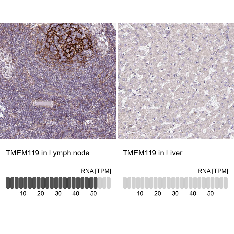
- Main image
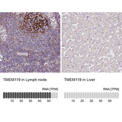
- Experimental details
- Immunohistochemistry analysis in human lymph node and liver tissues using HPA051870 antibody. Corresponding TMEM119 RNA-seq data are presented for the same tissues.
- Sample type
- Human
- Protocol
- Protocol