Explore
 Validate
 Learn
Learn Immunohistochemistry
Antibody data
- Antibody Data
- Antigen structure
- References [1]
- Comments [0]
- Validations
- Immunohistochemistry [1]
- Flow cytometry [1]
Submit
Validation data
Reference
Comment
Report error
- Product number
- AF7498 - Provider product page

- Provider
- R&D Systems
- Product name
- Human LRIG1 Antibody
- Antibody type
- Polyclonal
- Description
- Antigen Affinity-purified. Detects human LRIG1 in direct ELISAs. In direct ELISAs, approximately 10% cross-reactivity with recombinant mouse LRIG1 and and less than 1% cross-reactivity with recombinant human LRIG3 is observed.
- Reactivity
- Human
- Host
- Sheep
- Conjugate
- Unconjugated
- Antigen sequence
Q96JA1- Isotype
- IgG
- Vial size
- 100 ug
- Concentration
- LYOPH
- Storage
- Use a manual defrost freezer and avoid repeated freeze-thaw cycles. 12 months from date of receipt, -20 to -70 °C as supplied. 1 month, 2 to 8 °C under sterile conditions after reconstitution. 6 months, -20 to -70 °C under sterile conditions after reconstitution.
Submitted references USP8 modulates ubiquitination of LRIG1 for Met degradation.
Oh YM, Lee SB, Choi J, Suh HY, Shim S, Song YJ, Kim B, Lee JM, Oh SJ, Jeong Y, Cheong KH, Song PH, Kim KA
Scientific reports 2014 May 15;4:4980
Scientific reports 2014 May 15;4:4980
No comments: Submit comment
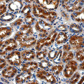
Supportive validation
- Submitted by
- R&D Systems (provider)
- Main image

- Experimental details
- LRIG1 in Human Kidney. LRIG1 was detected in immersion fixed paraffin-embedded sections of human kidney using Sheep Anti-Human LRIG1 Antigen Affinity-purified Polyclonal Antibody (Catalog # AF7498) at 10 µg/mL overnight at 4 °C. Before incubation with the primary antibody, tissue was subjected to heat-induced epitope retrieval using Antigen Retrieval Reagent-Basic (Catalog # CTS013). Tissue was stained using the Anti-Sheep HRP-DAB Cell & Tissue Staining Kit (brown; Catalog # CTS019) and counterstained with hematoxylin (blue). Specific staining was localized to the cytoplasm of epithelial cells in convoluted tubules. View our protocol for Chromogenic IHC Staining of Paraffin-embedded Tissue Sections.
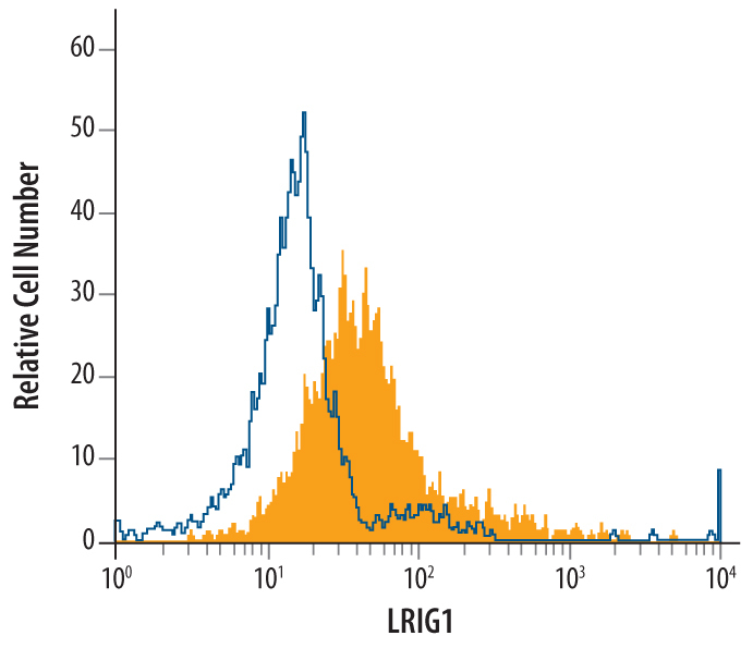
Supportive validation
- Submitted by
- R&D Systems (provider)
- Main image

- Experimental details
- Detection of LRIG1 in LNCaP Human Cell Line by Flow Cytometry. LNCaP human prostate cancer cell line was stained with Sheep Anti-Human LRIG1 Antigen Affinity-purified Polyclonal Antibody (Catalog # AF7498, filled histogram) or control antibody (Catalog # 5-001-A, open histogram), followed by Allophycocyanin-conjugated Anti-Sheep IgG Secondary Antibody (Catalog # F0127).