Explore
 Validate
 Learn
LearnHPA003156
antibody from Atlas Antibodies
Targeting: ATP6AP2
APT6M8-9, ATP6IP2, ATP6M8-9, M8-9, PRR, RENR
 Western blot
 Immunohistochemistry
Antibody data
- Antibody Data
- Antigen structure
- References [56]
- Comments [0]
- Validations
- Western blot [1]
Submit
Validation data
Reference
Comment
Report error
- Product number
- HPA003156 - Provider product page

- Provider
- Atlas Antibodies
- Proper citation
- Atlas Antibodies Cat#HPA003156, RRID:AB_1078245
- Product name
- Anti-ATP6AP2
- Antibody type
- Polyclonal
- Description
- Polyclonal Antibody against Human ATP6AP2, Gene description: ATPase, H+ transporting, lysosomal accessory protein 2, Alternative Gene Names: APT6M8-9, ATP6IP2, ATP6M8-9, M8-9, PRR, RENR, Validated applications: IHC, WB, Uniprot ID: O75787, Storage: Store at +4°C for short term storage. Long time storage is recommended at -20°C.
- Reactivity
- Human
- Host
- Rabbit
- Conjugate
- Unconjugated
- Isotype
- IgG
- Vial size
- 100 µl
- Concentration
- 0.2 mg/ml
- Storage
- Store at +4°C for short term storage. Long time storage is recommended at -20°C.
- Handling
- The antibody solution should be gently mixed before use.
Submitted references Renal Expression and Localization of the Receptor for (Pro)renin and Its Ligands in Rodent Models of Diabetes, Metabolic Syndrome, and Age-Dependent Focal and Segmental Glomerulosclerosis
OXGR1-Dependent (Pro)Renin Receptor Upregulation in Collecting Ducts of the Clipped Kidney Contributes to Na+ Balance in Goldblatt Hypertensive Mice
(Pro)renin receptor mediates tubular epithelial cell pyroptosis in diabetic kidney disease via DPP4-JNK pathway
Inhibition of Wnt/β-catenin signaling reduces renal fibrosis in murine glycogen storage disease type Ia
ATP6AP2, a regulator of LRP6/β-catenin protein trafficking, promotes Wnt/β-catenin signaling and bone formation in a cell type dependent manner
(Pro)Renin Receptor Decoy Peptide PRO20 Protects against Oxidative Renal Damage Induced by Advanced Oxidation Protein Products
(Pro)renin receptor promotes colorectal cancer progression through inhibiting the NEDD4L-mediated Wnt3 ubiquitination and modulating gut microbiota
Endothelial cell polarity and extracellular matrix composition require functional ATP6AP2 during developmental and pathological angiogenesis
Intramembrane client recognition potentiates the chaperone functions of calnexin
(Pro)renin Receptor Regulates Phosphate Homeostasis in Rats via Releasing Fibroblast Growth Factor-23
(Pro)renin receptor antagonist PRO20 attenuates nephrectomy‐induced nephropathy in rats via inhibition of intrarenal RAS and Wnt/β‐catenin signaling
Mutagenesis of the Cleavage Site of Pro Renin Receptor Abrogates Angiotensin II-Induced Hypertension in Mice
Site-1 Protease-Derived Soluble (Pro)Renin Receptor Contributes to Angiotensin II–Induced Hypertension in Mice
The (pro)renin receptor (ATP6ap2) facilitates receptor-mediated endocytosis and lysosomal function in the renal proximal tubule
The prorenin receptor and its soluble form contribute to lipid homeostasis
Knockout of Nephron ATP6AP2 Impairs Proximal Tubule Function and Prevents High-Fat Diet-Induced Obesity in Male Mice
(Pro)renin Receptor Is a Novel Independent Prognostic Marker in Invasive Urothelial Carcinoma of the Bladder
Diuretic Action of Apelin-13 Mediated by Inhibiting cAMP/PKA/sPRR Pathway
High glucose induces trafficking of prorenin receptor and stimulates profibrotic factors in the collecting duct
Novel regulation of renal gluconeogenesis by Atp6ap2 in response to high fat diet via PGC1-α/AKT-1 pathway
Cancer Stem Cells in Metastatic Head and Neck Cutaneous Squamous Cell Carcinoma Express Components of the Renin-Angiotensin System
(Pro)renin receptor decoy peptide PRO20 protects against adriamycin-induced nephropathy by targeting the intrarenal renin-angiotensin system
ELABELA antagonizes intrarenal renin-angiotensin system to lower blood pressure and protects against renal injury
Lipid droplet velocity is a microenvironmental sensor of aggressive tumors regulated by V-ATPase and PEDF
(Pro)renin receptor contributes to renal mitochondria dysfunction, apoptosis and fibrosis in diabetic mice
Expression of Components of the Renin-Angiotensin System in Pyogenic Granuloma
(Pro)renin receptor regulates lung development via the Wnt/β-catenin signaling pathway
(Pro)renin receptor contributes to pregnancy-induced sodium-water retention in rats via activation of intrarenal RAAS and α-ENaC
Pro-renin receptor suppresses mitochondrial biogenesis and function via AMPK/SIRT-1/ PGC-1α pathway in diabetic kidney
Radiation Nephropathy in a Nonhuman Primate Model of Partial-Body Irradiation With Minimal Bone Marrow Sparing—Part 2: Histopathology, Mediators, and Mechanisms
(Pro)renin Receptor Expression Increases throughout the Colorectal Adenoma—Adenocarcinoma Sequence and It Is Associated with Worse Colorectal Cancer Prognosis
(Pro)renin Receptor-Dependent Induction of Profibrotic Factors Is Mediated by COX-2/EP4/NOX-4/Smad Pathway in Collecting Duct Cells
Downregulated microRNA‑133a induces HUVECs injury: Potential role of the (pro) renin receptor in angiotensin�II‑dependent hypertension
Loss-of-function mutations in ATP6AP1 and ATP6AP2 in granular cell tumors
Essential role of ATP6AP2 enrichment in caveolae/lipid raft microdomains for the induction of neuronal differentiation of stem cells
Role of (pro)renin receptor in albumin overload-induced nephropathy in rats
Elevated (Pro)renin Receptor Expression Contributes to Maintaining Aerobic Metabolism in Growth Hormone Deficiency.
Atp6ap2 ablation in adult mice impairs viability through multiple organ deficiencies
Embryonic Stem Cell-Like Population in Dupuytren’s Disease Expresses Components of the Renin-Angiotensin System
(Pro)renin receptor activation increases profibrotic markers and fibroblast‐like phenotype through MAPK‐dependent ROS formation in mouse renal collecting duct cells
Cancer Stem Cells in Moderately Differentiated Lip Squamous Cell Carcinoma Express Components of the Renin–Angiotensin System
NF-κB-dependent upregulation of (pro)renin receptor mediates high-NaCl-induced apoptosis in mouse inner medullary collecting duct cells
Osteoblastic Lrp4 promotes osteoclastogenesis by regulating ATP release and adenosine-A2AR signaling
Mutations in the X-linked ATP6AP2 cause a glycosylation disorder with autophagic defects
Cancer stem cells in moderately differentiated oral tongue squamous cell carcinoma express components of the renin–angiotensin system
Human placental renin–angiotensin system in normotensive and pre‐eclamptic pregnancies at high altitude and after acute hypoxia–reoxygenation insult
Cancer Stem Cells in Moderately Differentiated Buccal Mucosal Squamous Cell Carcinoma Express Components of the Renin–Angiotensin System
Expression of the Components of the Renin–Angiotensin System in Venous Malformation
Vacuolar ATPase in Phagosome-Lysosome Fusion
Increased expression of (pro)renin receptor does not cause hypertension or cardiac and renal fibrosis in mice
Angiotensin II Increases the Expression of (Pro)Renin Receptor During Low-Salt Conditions
The (pro)renin receptor mediates constitutive PLZF-independent pro-proliferative effects which are inhibited by bafilomycin but not genistein
Renal medullary cyclooxygenase-2 and (pro)renin receptor expression during angiotensin II-dependent hypertension
Distinct Signal Transduction Pathways Downstream of the (P)RR Revealed by Microarray and ChIP-chip Analyses
Soluble Form of the (Pro)Renin Receptor Is Augmented in the Collecting Duct and Urine of Chronic Angiotensin II–Dependent Hypertensive Rats
Enhancement of renin and prorenin receptor in collecting duct of Cyp1a1-Ren2 rats may contribute to development and progression of malignant hypertension
Iacobini C, Vitale M, Sentinelli F, Haxhi J, Pugliese G, Menini S
International Journal of Molecular Sciences 2024;25(4):2217
International Journal of Molecular Sciences 2024;25(4):2217
OXGR1-Dependent (Pro)Renin Receptor Upregulation in Collecting Ducts of the Clipped Kidney Contributes to Na+ Balance in Goldblatt Hypertensive Mice
Cárdenas P, Nuñez-Allimant C, Silva K, Cid-Salinas C, León A, Vallotton Z, Lorca R, Oliveira L, Casarini D, Céspedes C, Prieto M, Gonzalez A
International Journal of Molecular Sciences 2024;25(18):10045
International Journal of Molecular Sciences 2024;25(18):10045
(Pro)renin receptor mediates tubular epithelial cell pyroptosis in diabetic kidney disease via DPP4-JNK pathway
Xie S, Song S, Liu S, Li Q, Zou W, Ke J, Wang C
Journal of Translational Medicine 2024;22(1)
Journal of Translational Medicine 2024;22(1)
Inhibition of Wnt/β-catenin signaling reduces renal fibrosis in murine glycogen storage disease type Ia
Lee C, Pratap K, Zhang L, Chen H, Gautam S, Arnaoutova I, Raghavankutty M, Starost M, Kahn M, Mansfield B, Chou J
Biochimica et Biophysica Acta (BBA) - Molecular Basis of Disease 2024;1870(1):166874
Biochimica et Biophysica Acta (BBA) - Molecular Basis of Disease 2024;1870(1):166874
ATP6AP2, a regulator of LRP6/β-catenin protein trafficking, promotes Wnt/β-catenin signaling and bone formation in a cell type dependent manner
Xiong L, Guo H, Pan J, Ren X, Lee D, Chen L, Mei L, Xiong W
Bone Research 2024;12(1)
Bone Research 2024;12(1)
(Pro)Renin Receptor Decoy Peptide PRO20 Protects against Oxidative Renal Damage Induced by Advanced Oxidation Protein Products
Fang H, Yang T, Zhou B, Li X
Molecules 2023;28(7):3017
Molecules 2023;28(7):3017
(Pro)renin receptor promotes colorectal cancer progression through inhibiting the NEDD4L-mediated Wnt3 ubiquitination and modulating gut microbiota
Wang J, Ding Y, Li D, Zhu N, Nishiyama A, Yuan Y
Cell Communication and Signaling 2023;21(1)
Cell Communication and Signaling 2023;21(1)
Endothelial cell polarity and extracellular matrix composition require functional ATP6AP2 during developmental and pathological angiogenesis
Patel N, K C R, Blanks A, Li Y, Prieto M, Meadows S
JCI Insight 2022;7(19)
JCI Insight 2022;7(19)
Intramembrane client recognition potentiates the chaperone functions of calnexin
Bloemeke N, Meighen‐Berger K, Hitzenberger M, Bach N, Parr M, Coelho J, Frishman D, Zacharias M, Sieber S, Feige M
The EMBO Journal 2022;41(24)
The EMBO Journal 2022;41(24)
(Pro)renin Receptor Regulates Phosphate Homeostasis in Rats via Releasing Fibroblast Growth Factor-23
Lu A, Pu M, Mo S, Su J, Hu J, Li C, Wang W, Yang T
Frontiers in Physiology 2022;13
Frontiers in Physiology 2022;13
(Pro)renin receptor antagonist PRO20 attenuates nephrectomy‐induced nephropathy in rats via inhibition of intrarenal RAS and Wnt/β‐catenin signaling
Wang Y, Wang Y, Xue K, Wang H, Zhou J, Gao F, Li C, Yang T, Fang H
Physiological Reports 2021;9(11)
Physiological Reports 2021;9(11)
Mutagenesis of the Cleavage Site of Pro Renin Receptor Abrogates Angiotensin II-Induced Hypertension in Mice
Wang F, Chen Y, Zou C, Luo R, Yang T
Hypertension 2021;78(1):115-127
Hypertension 2021;78(1):115-127
Site-1 Protease-Derived Soluble (Pro)Renin Receptor Contributes to Angiotensin II–Induced Hypertension in Mice
Feng Y, Peng K, Luo R, Wang F, Yang T
Hypertension 2021;77(2):405-416
Hypertension 2021;77(2):405-416
The (pro)renin receptor (ATP6ap2) facilitates receptor-mediated endocytosis and lysosomal function in the renal proximal tubule
Figueiredo M, Daryadel A, Sihn G, Müller D, Popova E, Rouselle A, Nguyen G, Bader M, Wagner C
Pflügers Archiv - European Journal of Physiology 2021;473(8):1229-1246
Pflügers Archiv - European Journal of Physiology 2021;473(8):1229-1246
The prorenin receptor and its soluble form contribute to lipid homeostasis
Gatineau E, Arthur G, Poupeau A, Nichols K, Spear B, Shelman N, Graf G, Temel R, Yiannikouris F
American Journal of Physiology-Endocrinology and Metabolism 2021;320(3):E609-E618
American Journal of Physiology-Endocrinology and Metabolism 2021;320(3):E609-E618
Knockout of Nephron ATP6AP2 Impairs Proximal Tubule Function and Prevents High-Fat Diet-Induced Obesity in Male Mice
Culver S, Akhtar S, Rountree-Jablin C, Keller S, Cathro H, Gildea J, Siragy H
Endocrinology 2021;162(12)
Endocrinology 2021;162(12)
(Pro)renin Receptor Is a Novel Independent Prognostic Marker in Invasive Urothelial Carcinoma of the Bladder
Larrinaga G, Calvete-Candenas J, Solano-Iturri J, Martín A, Pueyo A, Nunes-Xavier C, Pulido R, Dorado J, López J, Angulo J
Cancers 2021;13(22):5642
Cancers 2021;13(22):5642
Diuretic Action of Apelin-13 Mediated by Inhibiting cAMP/PKA/sPRR Pathway
Chen Y, Xu C, Hu J, Deng M, Qiu Q, Mo S, Du Y, Yang T
Frontiers in Physiology 2021;12
Frontiers in Physiology 2021;12
High glucose induces trafficking of prorenin receptor and stimulates profibrotic factors in the collecting duct
Gogulamudi V, Arita D, Bourgeois C, Jorgensen J, He J, Wimley W, Satou R, Gonzalez A, Prieto M
Scientific Reports 2021;11(1)
Scientific Reports 2021;11(1)
Novel regulation of renal gluconeogenesis by Atp6ap2 in response to high fat diet via PGC1-α/AKT-1 pathway
Akhtar S, Culver S, Siragy H
Scientific Reports 2021;11(1)
Scientific Reports 2021;11(1)
Cancer Stem Cells in Metastatic Head and Neck Cutaneous Squamous Cell Carcinoma Express Components of the Renin-Angiotensin System
Siljee S, Buchanan O, Brasch H, Bockett N, Patel J, Paterson E, Purdie G, Davis P, Itinteang T, Tan S
Cells 2021;10(2):243
Cells 2021;10(2):243
(Pro)renin receptor decoy peptide PRO20 protects against adriamycin-induced nephropathy by targeting the intrarenal renin-angiotensin system
Luo R, Yang K, Wang F, Xu C, Yang T
American Journal of Physiology-Renal Physiology 2020;319(5):F930-F940
American Journal of Physiology-Renal Physiology 2020;319(5):F930-F940
ELABELA antagonizes intrarenal renin-angiotensin system to lower blood pressure and protects against renal injury
Xu C, Wang F, Chen Y, Xie S, Sng D, Reversade B, Yang T
American Journal of Physiology-Renal Physiology 2020;318(5):F1122-F1135
American Journal of Physiology-Renal Physiology 2020;318(5):F1122-F1135
Lipid droplet velocity is a microenvironmental sensor of aggressive tumors regulated by V-ATPase and PEDF
Nardi F, Fitchev P, Brooks K, Franco O, Cheng K, Hayward S, Welte M, Crawford S
Laboratory Investigation 2019;99(12):1822-1834
Laboratory Investigation 2019;99(12):1822-1834
(Pro)renin receptor contributes to renal mitochondria dysfunction, apoptosis and fibrosis in diabetic mice
Li C, Matavelli L, Akhtar S, Siragy H
Scientific Reports 2019;9(1)
Scientific Reports 2019;9(1)
Expression of Components of the Renin-Angiotensin System in Pyogenic Granuloma
Papali'i-Curtin J, Brasch H, van Schaijik B, de Jongh J, Marsh R, Tan S, Itinteang T
Frontiers in Surgery 2019;6
Frontiers in Surgery 2019;6
(Pro)renin receptor regulates lung development via the Wnt/β-catenin signaling pathway
Liu J, Zhou Y, Liu Y, Li L, Chen Y, Liu Y, Feng Y, Yosypiv I, Song R, Peng H
American Journal of Physiology-Lung Cellular and Molecular Physiology 2019;317(2):L202-L211
American Journal of Physiology-Lung Cellular and Molecular Physiology 2019;317(2):L202-L211
(Pro)renin receptor contributes to pregnancy-induced sodium-water retention in rats via activation of intrarenal RAAS and α-ENaC
Fu Z, Hu J, Zhou L, Chen Y, Deng M, Liu X, Su J, Lu A, Fu X, Yang T
American Journal of Physiology-Renal Physiology 2019;316(3):F530-F538
American Journal of Physiology-Renal Physiology 2019;316(3):F530-F538
Pro-renin receptor suppresses mitochondrial biogenesis and function via AMPK/SIRT-1/ PGC-1α pathway in diabetic kidney
Sen U, Akhtar S, Siragy H
PLOS ONE 2019;14(12):e0225728
PLOS ONE 2019;14(12):e0225728
Radiation Nephropathy in a Nonhuman Primate Model of Partial-Body Irradiation With Minimal Bone Marrow Sparing—Part 2: Histopathology, Mediators, and Mechanisms
Parker G, Cohen E, Li N, Takayama K, Farese A, MacVittie T
Health Physics 2019;116(3):409-425
Health Physics 2019;116(3):409-425
(Pro)renin Receptor Expression Increases throughout the Colorectal Adenoma—Adenocarcinoma Sequence and It Is Associated with Worse Colorectal Cancer Prognosis
Beitia M, Solano-Iturri J, Errarte P, Calvete-Candenas J, Loizate A, Etxezarraga M, Sanz B, Larrinaga G
Cancers 2019;11(6):881
Cancers 2019;11(6):881
(Pro)renin Receptor-Dependent Induction of Profibrotic Factors Is Mediated by COX-2/EP4/NOX-4/Smad Pathway in Collecting Duct Cells
Reyes-Martinez C, Nguyen Q, Kassan M, Gonzalez A
Frontiers in Pharmacology 2019;10
Frontiers in Pharmacology 2019;10
Downregulated microRNA‑133a induces HUVECs injury: Potential role of the (pro) renin receptor in angiotensin�II‑dependent hypertension
Liu B, Lan M, Wei H, Zhang D, Liu J, Teng J
Molecular Medicine Reports 2019
Molecular Medicine Reports 2019
Loss-of-function mutations in ATP6AP1 and ATP6AP2 in granular cell tumors
Pareja F, Brandes A, Basili T, Selenica P, Geyer F, Fan D, Da Cruz Paula A, Kumar R, Brown D, Gularte-Mérida R, Alemar B, Bi R, Lim R, de Bruijn I, Fujisawa S, Gardner R, Feng E, Li A, da Silva E, Lozada J, Blecua P, Cohen-Gould L, Jungbluth A, Rakha E, Ellis I, Edelweiss M, Palazzo J, Norton L, Hollmann T, Edelweiss M, Rubin B, Weigelt B, Reis-Filho J
Nature Communications 2018;9(1)
Nature Communications 2018;9(1)
Essential role of ATP6AP2 enrichment in caveolae/lipid raft microdomains for the induction of neuronal differentiation of stem cells
Makdissy N, Haddad K, AlBacha J, Chaker D, Ismail B, Azar A, Oreibi G, Ayoub D, Achkar I, Quilliot D, Fajloun Z
Stem Cell Research & Therapy 2018;9(1)
Stem Cell Research & Therapy 2018;9(1)
Role of (pro)renin receptor in albumin overload-induced nephropathy in rats
Fang H, Deng M, Zhang L, Lu A, Su J, Xu C, Zhou L, Wang L, Ou J, Wang W, Yang T
American Journal of Physiology-Renal Physiology 2018;315(6):F1759-F1768
American Journal of Physiology-Renal Physiology 2018;315(6):F1759-F1768
Elevated (Pro)renin Receptor Expression Contributes to Maintaining Aerobic Metabolism in Growth Hormone Deficiency.
Seki Y, Yatabe M, Suda C, Morimoto S, Ichihara A
Journal of the Endocrine Society 2018 Mar 1;2(3):252-265
Journal of the Endocrine Society 2018 Mar 1;2(3):252-265
Atp6ap2 ablation in adult mice impairs viability through multiple organ deficiencies
Wendling O, Champy M, Jaubert S, Pavlovic G, Dubos A, Lindner L, Jacobs H, Mark M, Combe R, Da Cruz I, Luche H, Mudgett J, Rosahl T, Sorg T, Malissen M, Reilly P, Hérault Y
Scientific Reports 2017;7(1)
Scientific Reports 2017;7(1)
Embryonic Stem Cell-Like Population in Dupuytren’s Disease Expresses Components of the Renin-Angiotensin System
On N, Koh S, Brasch H, Dunne J, Armstrong J, Tan S, Itinteang T
Plastic and Reconstructive Surgery - Global Open 2017;5(7):e1422
Plastic and Reconstructive Surgery - Global Open 2017;5(7):e1422
(Pro)renin receptor activation increases profibrotic markers and fibroblast‐like phenotype through MAPK‐dependent ROS formation in mouse renal collecting duct cells
Gonzalez A, Zamora L, Reyes‐Martinez C, Salinas‐Parra N, Roldan N, Cuevas C, Figueroa S, Gonzalez‐Vergara A, Prieto M
Clinical and Experimental Pharmacology and Physiology 2017;44(11):1134-1144
Clinical and Experimental Pharmacology and Physiology 2017;44(11):1134-1144
Cancer Stem Cells in Moderately Differentiated Lip Squamous Cell Carcinoma Express Components of the Renin–Angiotensin System
Ram R, Brasch H, Dunne J, Davis P, Tan S, Itinteang T
Frontiers in Surgery 2017;4
Frontiers in Surgery 2017;4
NF-κB-dependent upregulation of (pro)renin receptor mediates high-NaCl-induced apoptosis in mouse inner medullary collecting duct cells
Su J, Liu X, Xu C, Lu X, Wang F, Fang H, Lu A, Qiu Q, Li C, Yang T
American Journal of Physiology-Cell Physiology 2017;313(6):C612-C620
American Journal of Physiology-Cell Physiology 2017;313(6):C612-C620
Osteoblastic Lrp4 promotes osteoclastogenesis by regulating ATP release and adenosine-A2AR signaling
Xiong L, Jung J, Guo H, Pan J, Sun X, Mei L, Xiong W
Journal of Cell Biology 2017;216(3):761-778
Journal of Cell Biology 2017;216(3):761-778
Mutations in the X-linked ATP6AP2 cause a glycosylation disorder with autophagic defects
Rujano M, Cannata Serio M, Panasyuk G, Péanne R, Reunert J, Rymen D, Hauser V, Park J, Freisinger P, Souche E, Guida M, Maier E, Wada Y, Jäger S, Krogan N, Kretz O, Nobre S, Garcia P, Quelhas D, Bird T, Raskind W, Schwake M, Duvet S, Foulquier F, Matthijs G, Marquardt T, Simons M
Journal of Experimental Medicine 2017;214(12):3707-3729
Journal of Experimental Medicine 2017;214(12):3707-3729
Cancer stem cells in moderately differentiated oral tongue squamous cell carcinoma express components of the renin–angiotensin system
Itinteang T, Dunne J, Chibnall A, Brasch H, Davis P, Tan S
Journal of Clinical Pathology 2016;69(10):942-945
Journal of Clinical Pathology 2016;69(10):942-945
Human placental renin–angiotensin system in normotensive and pre‐eclamptic pregnancies at high altitude and after acute hypoxia–reoxygenation insult
Kurlak L, Mistry H, Cindrova‐Davies T, Burton G, Pipkin F
The Journal of Physiology 2016;594(5):1327-1340
The Journal of Physiology 2016;594(5):1327-1340
Cancer Stem Cells in Moderately Differentiated Buccal Mucosal Squamous Cell Carcinoma Express Components of the Renin–Angiotensin System
Featherston T, Yu H, Dunne J, Chibnall A, Brasch H, Davis P, Tan S, Itinteang T
Frontiers in Surgery 2016;3
Frontiers in Surgery 2016;3
Expression of the Components of the Renin–Angiotensin System in Venous Malformation
Siljee S, Keane E, Marsh R, Brasch H, Tan S, Itinteang T
Frontiers in Surgery 2016;3
Frontiers in Surgery 2016;3
Vacuolar ATPase in Phagosome-Lysosome Fusion
Kissing S, Hermsen C, Repnik U, Nesset C, von Bargen K, Griffiths G, Ichihara A, Lee B, Schwake M, De Brabander J, Haas A, Saftig P
Journal of Biological Chemistry 2015;290(22):14166-14180
Journal of Biological Chemistry 2015;290(22):14166-14180
Increased expression of (pro)renin receptor does not cause hypertension or cardiac and renal fibrosis in mice
Rosendahl A, Niemann G, Lange S, Ahadzadeh E, Krebs C, Contrepas A, van Goor H, Wiech T, Bader M, Schwake M, Peters J, Stahl R, Nguyen G, Wenzel U
Laboratory Investigation 2014;94(8):863-872
Laboratory Investigation 2014;94(8):863-872
Angiotensin II Increases the Expression of (Pro)Renin Receptor During Low-Salt Conditions
Gonzalez A, Womack J, Liu L, Prieto M, Seth D
The American Journal of the Medical Sciences 2014;348(5):416-422
The American Journal of the Medical Sciences 2014;348(5):416-422
The (pro)renin receptor mediates constitutive PLZF-independent pro-proliferative effects which are inhibited by bafilomycin but not genistein
KIRSCH S, SCHREZENMEIER E, KLARE S, ZAADE D, SEIDEL K, SCHMITZ J, BERNHARD S, LAUER D, SLACK M, GOLDIN-LANG P, UNGER T, ZOLLMANN F, FUNKE-KAISER H
International Journal of Molecular Medicine 2014;33(4):795-808
International Journal of Molecular Medicine 2014;33(4):795-808
Renal medullary cyclooxygenase-2 and (pro)renin receptor expression during angiotensin II-dependent hypertension
Gonzalez A, Green T, Luffman C, Bourgeois C, Gabriel Navar L, Prieto M
American Journal of Physiology-Renal Physiology 2014;307(8):F962-F970
American Journal of Physiology-Renal Physiology 2014;307(8):F962-F970
Distinct Signal Transduction Pathways Downstream of the (P)RR Revealed by Microarray and ChIP-chip Analyses
Datta P, Zaade D, Schmitz J, Benke E, Klare S, Seidel K, Kirsch S, Goldin-Lang P, Zollmann F, Unger T, Funke-Kaiser H
PLoS ONE 2013;8(3):e57674
PLoS ONE 2013;8(3):e57674
Soluble Form of the (Pro)Renin Receptor Is Augmented in the Collecting Duct and Urine of Chronic Angiotensin II–Dependent Hypertensive Rats
Gonzalez A, Lara L, Luffman C, Seth D, Prieto M
Hypertension 2011;57(4):859-864
Hypertension 2011;57(4):859-864
Enhancement of renin and prorenin receptor in collecting duct of Cyp1a1-Ren2 rats may contribute to development and progression of malignant hypertension
Prieto M, Williams D, Liu L, Kavanagh K, Mullins J, Mitchell K
American Journal of Physiology-Renal Physiology 2011;300(2):F581-F588
American Journal of Physiology-Renal Physiology 2011;300(2):F581-F588
No comments: Submit comment
Enhanced validation
- Submitted by
- Atlas Antibodies (provider)
- Enhanced method
- Genetic validation
- Main image

- Experimental details
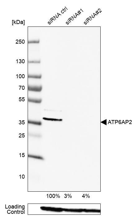
- Western blot analysis in HEK293 cells transfected with control siRNA, target specific siRNA probe #1 and #2, using Anti-ATP6AP2 antibody. Remaining relative intensity is presented. Loading control: Anti-PPIB.
- Sample type
- Human
- Protocol
- Protocol