 Explore
 Validate
 Learn
Learn Western blot
Antibody data
- Antibody Data
- Antigen structure
- References [2]
- Comments [0]
- Validations
- Western blot [2]
- Other assay [4]
Submit
Validation data
Reference
Comment
Report error
- Product number
- PA5-76821 - Provider product page

- Provider
- Invitrogen Antibodies
- Product name
- Biglycan Polyclonal Antibody
- Antibody type
- Polyclonal
- Antigen
- Recombinant full-length protein
- Description
- The antibody was affinity-purified from rabbit antiserum by affinity-chromatography using epitope-specific immunogen and the purity is > 95% (by SDS-PAGE).
- Reactivity
- Human, Mouse, Rat
- Host
- Rabbit
- Isotype
- IgG
- Vial size
- 100 µL
- Concentration
- 1 mg/mL
- Storage
- Store at 4°C short term. For long term storage, store at -20°C, avoiding freeze/thaw cycles.
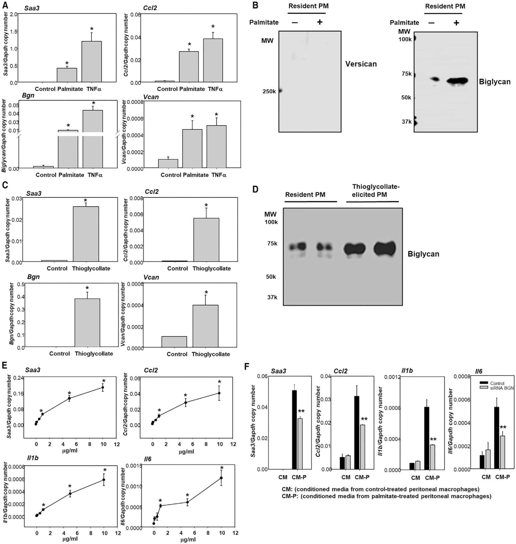
Submitted references Serum amyloid A-containing HDL binds adipocyte-derived versican and macrophage-derived biglycan, reducing its antiinflammatory properties.
Adipocyte-Derived Versican and Macrophage-Derived Biglycan Control Adipose Tissue Inflammation in Obesity.
Han CY, Kang I, Omer M, Wang S, Wietecha T, Wight TN, Chait A
JCI insight 2020 Sep 24;5(20)
JCI insight 2020 Sep 24;5(20)
Adipocyte-Derived Versican and Macrophage-Derived Biglycan Control Adipose Tissue Inflammation in Obesity.
Han CY, Kang I, Harten IA, Gebe JA, Chan CK, Omer M, Alonge KM, den Hartigh LJ, Gomes Kjerulf D, Goodspeed L, Subramanian S, Wang S, Kim F, Birk DE, Wight TN, Chait A
Cell reports 2020 Jun 30;31(13):107818
Cell reports 2020 Jun 30;31(13):107818
No comments: Submit comment
Supportive validation
- Submitted by
- Invitrogen Antibodies (provider)
- Main image

- Experimental details
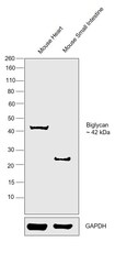
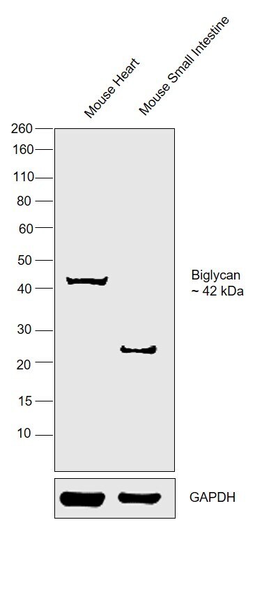
- Western blot analysis of Biglycan. Samples were incubated with Biglycan polyclonal antibody (Product # PA5-76821).

- Submitted by
- Invitrogen Antibodies (provider)
- Main image
- Experimental details
- Western blot was performed using Anti-Biglycan Polyclonal Antibody (Product # PA5-76821) and a 42 kDa bands corresponding to Biglycan was observed in Mouse Heart but not in Mouse Skeletal Muscle. An uncharacterized bands of ~23 kDa was observed in negative cell line. Tissue extracts (30 µg lysate) of Mouse Heart (Lane 1) and Mouse Small Intestine (Lane 2) were electrophoresed using Novex® NuPAGE® 4-12 % Bis-Tris gel (Product # NP0321BOX). Resolved proteins were then transferred onto a nitrocellulose membrane (Product # IB23001) by iBlot® 2 Dry Blotting System (Product # IB21001). The blot was probed with the primary antibody (1:1000 dilution) and detected by chemiluminescence with Goat anti-Rabbit IgG (H+L), Superclonal™ Recombinant Secondary Antibody, HRP (Product # A27036), using the iBright FL 1000 (Product # A32752). Chemiluminescent detection was performed using Novex® ECL Chemiluminescent Substrate Reagent Kit (Product # WP20005).
Supportive validation
- Submitted by
- Invitrogen Antibodies (provider)
- Main image
- Experimental details
- NULL
- Submitted by
- Invitrogen Antibodies (provider)
- Main image

- Experimental details
- NULL
- Submitted by
- Invitrogen Antibodies (provider)
- Main image
- Experimental details
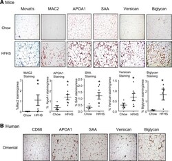
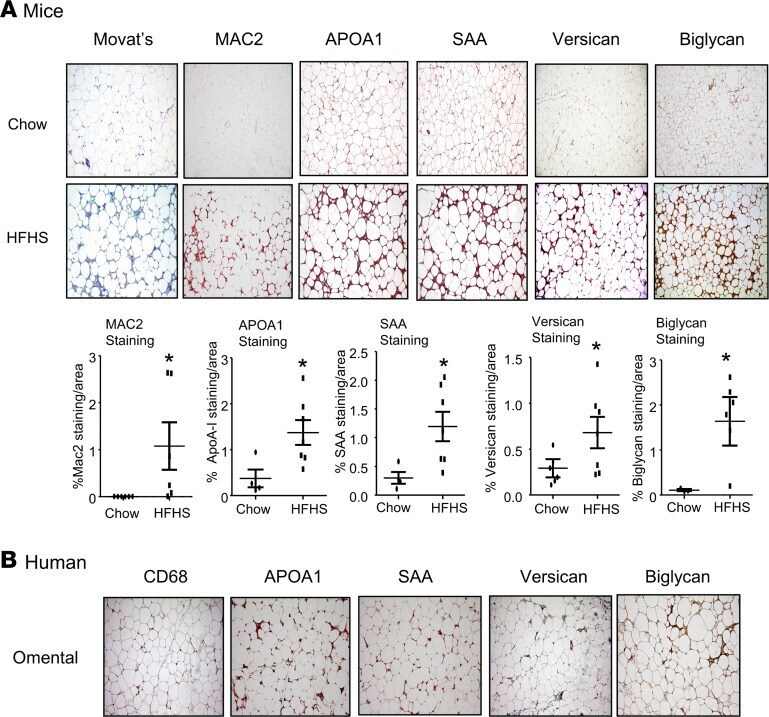
- Figure 1 Immunohistochemical staining of versican and biglycan shows a similar distribution with the staining of SAA and APOA1 in the epididymal white adipose tissue from mice fed an HFHS diet and in the omental fat tissue from human obese subjects undergoing gastric bypass surgery. ( A ) Mice were fed a chow or HFHS diet for 16 weeks, after which adipose tissue was obtained and immunostained with the antibodies shown. Tissues were photographed using microscopy (original magnification, x60). Quantitation of the immunostaining is shown in the lower panel ( n = 5-7, mean +- SEM). * P < 0.001 vs. chow. ( B ) Omental fat was obtained from gastric bypass patients and immunostained with the antibodies shown. Tissues were photographed using microscopy (original magnification, x60). The pictures of MAC2, CD68, versican, and biglycan staining are from our previous publication (); APOA1 and SAA staining were performed on adjacent sections.
- Submitted by
- Invitrogen Antibodies (provider)
- Main image
- Experimental details
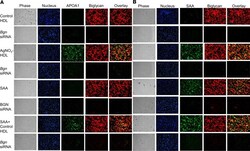
- Figure 5 HDL isolated from AgNO 3 -injected mice colocalizes with biglycan at the cell surface of peritoneal macrophages. HDL from PBS- and AgNO 3 -injected C57BL/6 mice was isolated. After exposure to these HDL preparations (50 mug protein/mL) for 6 hours, TG-elicited peritoneal macrophages from Saa3 -/- were fixed in 2% formalin for 5 minutes ( A and B ). After extensive washing, ( A ) APOA1 and biglycan were stained using anti-biglycan (red) and anti-APOA1 (green) antibodies, or ( B ) SAA and biglycan were stained using anti-biglycan (red) and ant-SAA (green) antibodies and photographed by fluorescence microscopy (Nikon Eclipse 80i, original magnification, x200).
