 Explore
 Validate
 Learn
Learn Western blot
 Immunocytochemistry
Antibody data
- Antibody Data
- Antigen structure
- References [10]
- Comments [0]
- Validations
- Western blot [1]
- Immunocytochemistry [1]
- Immunohistochemistry [1]
Submit
Validation data
Reference
Comment
Report error
- Product number
- HPA003157 - Provider product page

- Provider
- Atlas Antibodies
- Proper citation
- Atlas Antibodies Cat#HPA003157, RRID:AB_1078282
- Product name
- Anti-BGN
- Antibody type
- Polyclonal
- Description
- Polyclonal Antibody against Human BGN, Gene description: biglycan, Alternative Gene Names: DSPG1, SLRR1A, Validated applications: ICC, IHC, WB, Uniprot ID: P21810, Storage: Store at +4°C for short term storage. Long time storage is recommended at -20°C.
- Reactivity
- Human
- Host
- Rabbit
- Conjugate
- Unconjugated
- Isotype
- IgG
- Vial size
- 100 µl
- Concentration
- 0.1 mg/ml
- Storage
- Store at +4°C for short term storage. Long time storage is recommended at -20°C.
- Handling
- The antibody solution should be gently mixed before use.
Submitted references Deep learning neural network image analysis of immunohistochemical protein expression reveals a significantly reduced expression of biglycan in breast cancer
The Extracellular Small Leucine-Rich Proteoglycan Biglycan Is a Key Player in Gastric Cancer Aggressiveness
Toll-like receptor 4 signaling activates ERG function in prostate cancer and provides a therapeutic target.
Single-cell mapping reveals new markers and functions of lymphatic endothelial cells in lymph nodes
Early extracellular matrix changes are associated with later development of bronchiolitis obliterans syndrome after lung transplantation.
Up-regulation of Biglycan is Associated with Poor Prognosis and PTEN Deletion in Patients with Prostate Cancer
Sterile inflammation as a factor in human male infertility: Involvement of Toll like receptor 2, biglycan and peritubular cells
Blood biomarkers for the non-invasive diagnosis of endometriosis
Differential regulation of extracellular matrix protein expression in carcinoma-associated fibroblasts by TGF-β1 regulates cancer cell spreading but not adhesion.
Immunofluorescence and fluorescent-protein tagging show high correlation for protein localization in mammalian cells
Pavao M, Thiesen A, Mielczarski B, Savaris R
PLOS ONE 2023;18(3):e0282176
PLOS ONE 2023;18(3):e0282176
The Extracellular Small Leucine-Rich Proteoglycan Biglycan Is a Key Player in Gastric Cancer Aggressiveness
Pinto F, Santos-Ferreira L, Pinto M, Gomes C, Reis C
Cancers 2021;13(6):1330
Cancers 2021;13(6):1330
Toll-like receptor 4 signaling activates ERG function in prostate cancer and provides a therapeutic target.
Greulich BM, Plotnik JP, Jerde TJ, Hollenhorst PC
NAR cancer 2021 Mar;3(1):zcaa046
NAR cancer 2021 Mar;3(1):zcaa046
Single-cell mapping reveals new markers and functions of lymphatic endothelial cells in lymph nodes
Cyster J, Fujimoto N, He Y, D’Addio M, Tacconi C, Detmar M, Dieterich L
PLOS Biology 2020;18(4):e3000704
PLOS Biology 2020;18(4):e3000704
Early extracellular matrix changes are associated with later development of bronchiolitis obliterans syndrome after lung transplantation.
Müller C, Andersson-Sjöland A, Schultz HH, Eriksson LT, Andersen CB, Iversen M, Westergren-Thorsson G
BMJ open respiratory research 2017;4(1):e000177
BMJ open respiratory research 2017;4(1):e000177
Up-regulation of Biglycan is Associated with Poor Prognosis and PTEN Deletion in Patients with Prostate Cancer
Jacobsen F, Kraft J, Schroeder C, Hube-Magg C, Kluth M, Lang D, Simon R, Sauter G, Izbicki J, Clauditz T, Luebke A, Hinsch A, Wilczak W, Wittmer C, Büscheck F, Höflmayer D, Minner S, Tsourlakis M, Huland H, Graefen M, Budäus L, Thederan I, Salomon G, Schlomm T, Melling N
Neoplasia 2017;19(9):707-715
Neoplasia 2017;19(9):707-715
Sterile inflammation as a factor in human male infertility: Involvement of Toll like receptor 2, biglycan and peritubular cells
Mayer C, Adam M, Glashauser L, Dietrich K, Schwarzer J, Köhn F, Strauss L, Welter H, Poutanen M, Mayerhofer A
Scientific Reports 2016;6(1)
Scientific Reports 2016;6(1)
Blood biomarkers for the non-invasive diagnosis of endometriosis
Nisenblat V, Bossuyt P, Shaikh R, Farquhar C, Jordan V, Scheffers C, Mol B, Johnson N, Hull M
Cochrane Database of Systematic Reviews 2016;2016(5)
Cochrane Database of Systematic Reviews 2016;2016(5)
Differential regulation of extracellular matrix protein expression in carcinoma-associated fibroblasts by TGF-β1 regulates cancer cell spreading but not adhesion.
Van Bockstal M, Lambein K, Van Gele M, De Vlieghere E, Limame R, Braems G, Van den Broecke R, Cocquyt V, Denys H, Bracke M, Libbrecht L, De Wever O
Oncoscience 2014;1(10):634-48
Oncoscience 2014;1(10):634-48
Immunofluorescence and fluorescent-protein tagging show high correlation for protein localization in mammalian cells
Stadler C, Rexhepaj E, Singan V, Murphy R, Pepperkok R, Uhlén M, Simpson J, Lundberg E
Nature Methods 2013;10(4):315-323
Nature Methods 2013;10(4):315-323
No comments: Submit comment
Enhanced validation
- Submitted by
- Atlas Antibodies (provider)
- Enhanced method
- Recombinant expression validation
- Main image
- Experimental details
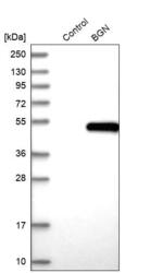
- Western blot analysis in control (vector only transfected HEK293T lysate) and BGN over-expression lysate (Co-expressed with a C-terminal myc-DDK tag (~3.1 kDa) in mammalian HEK293T cells, LY400642).
- Sample type
- Human
- Protocol
- Protocol

Supportive validation
- Submitted by
- Atlas Antibodies (provider)
- Main image
- Experimental details
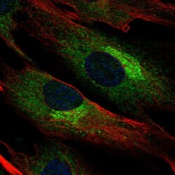
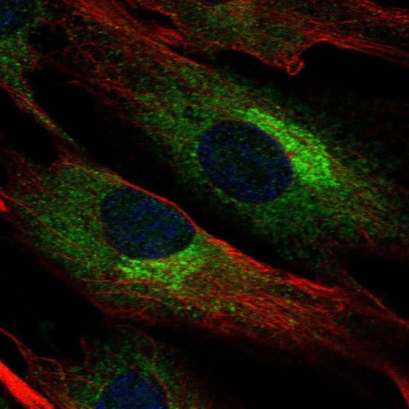
- Immunofluorescent staining of human cell line BJ shows localization to endoplasmic reticulum & the Golgi apparatus.
- Sample type
- Human
Supportive validation
- Submitted by
- Atlas Antibodies (provider)
- Enhanced method
- Orthogonal validation
- Main image
- Experimental details
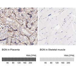
- Immunohistochemistry analysis in human placenta and skeletal muscle tissues using HPA003157 antibody. Corresponding BGN RNA-seq data are presented for the same tissues.
- Sample type
- Human
- Protocol
- Protocol
