Explore
 Validate
 Learn
Learn Western blot
 Immunohistochemistry
Antibody data
- Antibody Data
- Antigen structure
- References [2]
- Comments [0]
- Validations
- Immunohistochemistry [2]
- Other assay [1]
Submit
Validation data
Reference
Comment
Report error
- Product number
- PA5-79128 - Provider product page

- Provider
- Invitrogen Antibodies
- Product name
- CYP27B1 Polyclonal Antibody
- Antibody type
- Polyclonal
- Antigen
- Synthetic peptide
- Description
- Reconstitute with 0.2 mL of distilled water to yield a concentration of 500 µg/mL. Positive Control - WB: human Hela whole cell, human Caco-2 whole cell, rat heart tissue, rat brain tissue, mouse heart tissue, mouse RAW2647 whole cell. IHC: rat kidney tissue, human kidney cancer tissue.
- Reactivity
- Human, Mouse, Rat
- Host
- Rabbit
- Isotype
- IgG
- Vial size
- 100 μg
- Concentration
- 500 μg/mL
- Storage
- -20°C
Submitted references Dietary branched-chain amino acids modulate the dynamics of calcium absorption and reabsorption in protein-restricted pigs.
Vitamin D(3) Metabolic Enzymes in the Porcine Uterus: Expression, Localization and Autoregulation by 1,25(OH)(2)D(3) In Vitro.
Habibi M, Shili CN, Sutton J, Goodarzi P, Pezeshki A
Journal of animal science and biotechnology 2022 Feb 10;13(1):15
Journal of animal science and biotechnology 2022 Feb 10;13(1):15
Vitamin D(3) Metabolic Enzymes in the Porcine Uterus: Expression, Localization and Autoregulation by 1,25(OH)(2)D(3) In Vitro.
Grzesiak M, Kaminska K, Bodzioch A, Drzewiecka EM, Franczak A, Knapczyk-Stwora K
International journal of molecular sciences 2022 Apr 2;23(7)
International journal of molecular sciences 2022 Apr 2;23(7)
No comments: Submit comment
Supportive validation
- Submitted by
- Invitrogen Antibodies (provider)
- Main image
- Experimental details
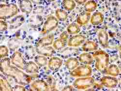
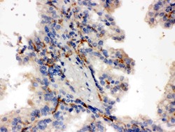
- Immunohistochemistry analysis of CYP27B1 on paraffin-embedded rat kidney tissue. Sample was incubated with CYP27B1 polyclonal antibody (Product# PA5-79128).
- Submitted by
- Invitrogen Antibodies (provider)
- Main image
- Experimental details
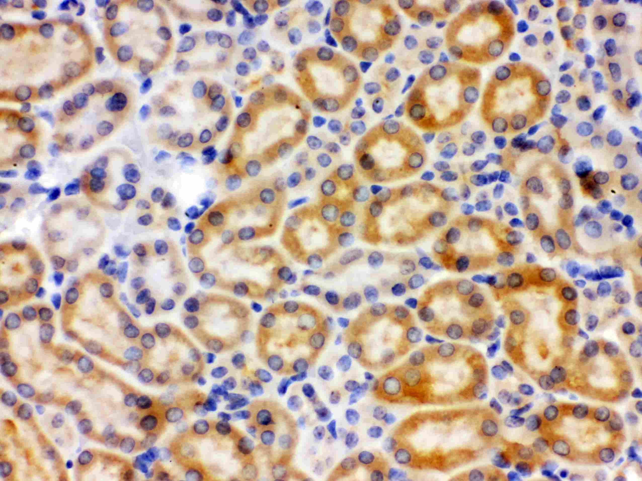
- Immunohistochemistry analysis of CYP27B1 on paraffin-embedded human kidney cancer tissue. Sample was incubated with CYP27B1 polyclonal antibody (Product# PA5-79128).

Supportive validation
- Submitted by
- Invitrogen Antibodies (provider)
- Main image
- Experimental details
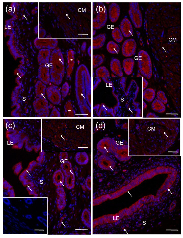
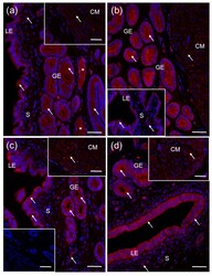
- Representative micrographs of CYP27B1 immunofluorescent localization within porcine uterus on days 2-5 ( a ), 10-12 ( b ), 15-16 ( c ) and 18-20 ( d ) of the estrous cycle. Immunoreactive proteins were visualized using a Cy3 detection system (red). Nuclei were counterstained with DAPI (blue). Positive signal ( arrows ) was found in the cytoplasm of luminal (LE) and glandular (GE) epithelial cells of the endometrium, and myocytes within whole myometrial layer (only a circular myometrium (CM) is presented herein). Negative control ( c lower inset) was obtained by the replacement of primary antibody by non-immune rabbit IgG. S, stroma; asterisks (*), blood vessels. Bar = 50 um.