Explore
 Validate
 Learn
Learn Western blot
Antibody data
- Antibody Data
- Antigen structure
- References [2]
- Comments [0]
- Validations
- Western blot [2]
- Immunohistochemistry [1]
- Flow cytometry [2]
- Other assay [1]
Submit
Validation data
Reference
Comment
Report error
- Product number
- PA5-26065 - Provider product page

- Provider
- Invitrogen Antibodies
- Product name
- CYP27B1 Polyclonal Antibody
- Antibody type
- Polyclonal
- Antigen
- Synthetic peptide
- Reactivity
- Human, Rat
- Host
- Rabbit
- Isotype
- IgG
- Vial size
- 200 μL
- Concentration
- 0.5 mg/mL
- Storage
- Store at 4°C short term. For long term storage, store at -20°C, avoiding freeze/thaw cycles.
Submitted references Hypercalcemia Owing to Overproduction of 1,25-Dihydroxyvitamin D(3) in Fetal Lung Adenocarcinoma: Case Report.
Mast cells express CYP27A1 and CYP27B1 in epithelial skin cancers and psoriasis.
Saito T, Ishida M, Kusabe M, Utsumi T, Maru N, Matsui H, Taniguchi Y, Kurata T, Kurokawa H, Imada T, Tsuta K, Tsukaguchi H, Murakawa T
JTO clinical and research reports 2021 Aug;2(8):100204
JTO clinical and research reports 2021 Aug;2(8):100204
Mast cells express CYP27A1 and CYP27B1 in epithelial skin cancers and psoriasis.
Kaukinen A, Pelkonen J, Harvima IT
European journal of dermatology : EJD 2015 Nov-Dec;25(6):548-55
European journal of dermatology : EJD 2015 Nov-Dec;25(6):548-55
No comments: Submit comment
Supportive validation
- Submitted by
- Invitrogen Antibodies (provider)
- Main image
- Experimental details
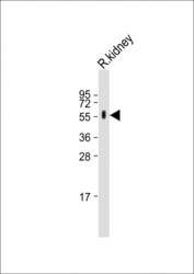
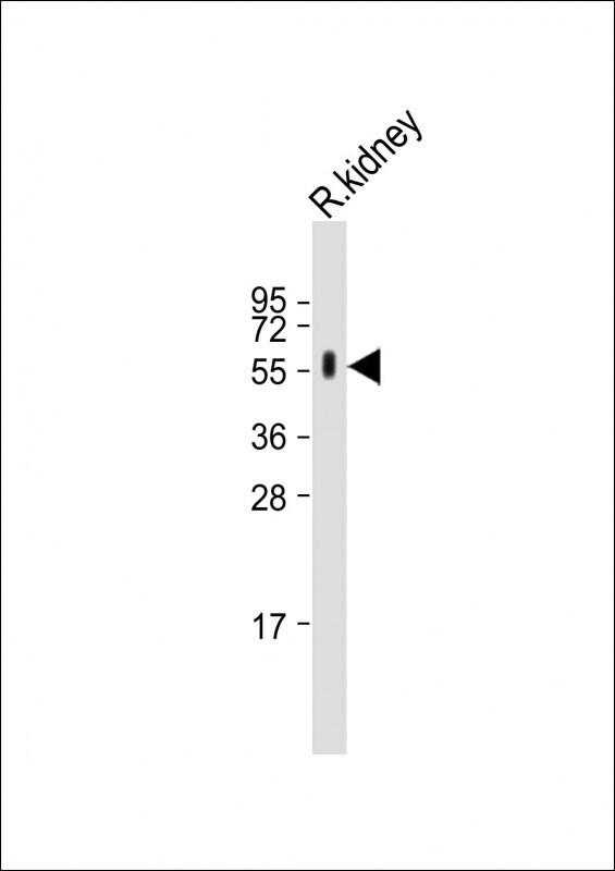
- Western blot analysis of CYP27B1 in Rat kidney whole tissue lysate. Samples were incubated with CYP27B1 polyclonal antibody (Product # PA5-26065) using a dilution of 1:2,000 followed by Goat Anti-Rabbit IgG, (H+L), Peroxidase conjugated at a dilution of 1:10,000. Lysates/proteins: 20 µg per lane. Predicted band size: 57 kDa. Blocking/Dilution buffer: 5% NFDM/TBST.
- Submitted by
- Invitrogen Antibodies (provider)
- Main image

- Experimental details
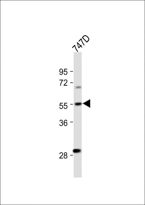
- Western blot analysis of CYP27B1 in T47D whole cell lysate. Samples were incubated with CYP27B1 polyclonal antibody (Product # PA5-26065) using a dilution of 1:500 followed by Goat Anti-Rabbit IgG, (H+L), Peroxidase conjugated at a dilution of 1:10,000. Lysates/proteins: 20 µg per lane. Predicted band size: 57 kDa. Blocking/Dilution buffer: 5% NFDM/TBST.
Supportive validation
- Submitted by
- Invitrogen Antibodies (provider)
- Main image
- Experimental details
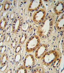
- Immunohistochemistry analysis of CYP27B1 in formalin fixed and paraffin embedded human kidney tissue. Samples were incubated with CYP27B1 polyclonal antibody (Product # PA5-26065) followed by peroxidase conjugation of the secondary antibody and DAB staining. This data demonstrates the use of this antibody for immunohistochemistry. Clinical relevance has not been evaluated.

Supportive validation
- Submitted by
- Invitrogen Antibodies (provider)
- Main image

- Experimental details
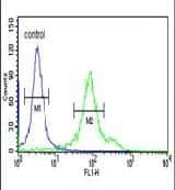
- Flow cytometry analysis of 293 cells using a CYP27B1 polyclonal antibody (Product # PA5-26065) (right) compared to a negative control cell (left) at a dilution of 1:10-50, followed by a FITC-conjugated goat anti-rabbit antibody
- Submitted by
- Invitrogen Antibodies (provider)
- Main image
- Experimental details
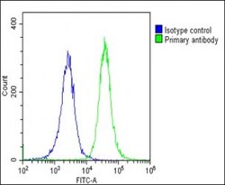
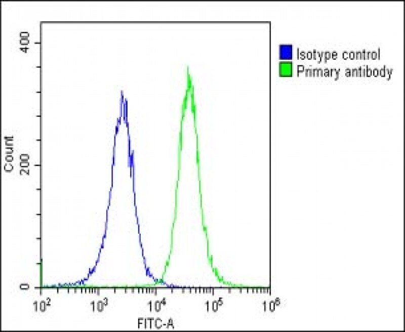
- Flow cytometry of (overlay histogram) of CYP27B1 in Hela cells (green line). Samples were incubated with CYP27B1 polyclonal antibody (Product # PA5-26065) using a dilution of 1:25 dilution for 60 min at 37°C followed by Goat-Anti-Rabbit IgG, DyLight® 488 Conjugated Highly Cross-Adsorbed at 1:200 dilution for 40 min at 37°C. The cells were fixed with 2% paraformaldehyde (10 min) and then permeabilized with 90% methanol for 10 min. The cells were then incubated in 2% bovine serum albumin to block non-specific protein-protein interactions followed by the primary antibody. Isotype control antibody (blue line) was rabbit IgG1 (1 μg/1x10^6 cells) used under the same conditions. Acquisition of >10, 000 events was performed.
Supportive validation
- Submitted by
- Invitrogen Antibodies (provider)
- Main image
- Experimental details
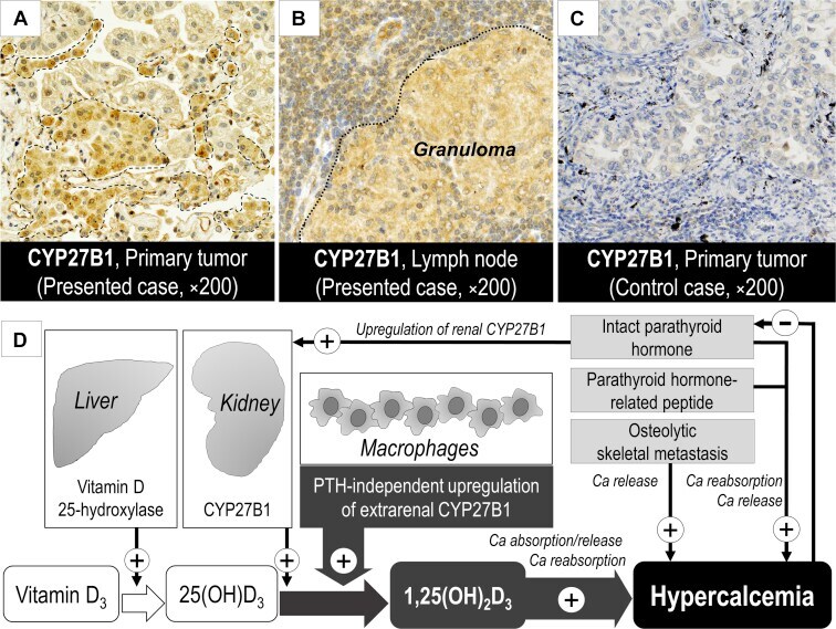
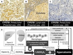
- Figure 3 Immunohistochemical staining of CYP27B1 and schematic presentation of the pathophysiology of vitamin D 3 -mediated hypercalcemia. Immunostaining for CYP27B1 (PA5-26065, Thermo Fisher Scientific, Rockford, IL) revealed a strong positivity in alveolar macrophages (surrounded by broken line), ( A ) weak positivity in tumor cells, and ( B ) strong positivity in macrophages in granulomas within the dissected lymph nodes (below the dotted line). ( C ) Another control case with pulmonary adenocarcinoma revealed CYP27B1 negativity. ( D ) In the presented case, hypercalcemia might be induced by extrarenal CYP27B1-mediated 1,25(OH) 2 D 3 overproduction (thick gray arrows). 1,25(OH) 2 D 3 , 1,25-dihydroxyvitaminD 3 ; 25(OH)D 3 , 25-hydroxyvitamin D 3 ; Ca, calcium.