Explore
 Validate
 Learn
Learn Western blot
Antibody data
- Antibody Data
- Antigen structure
- References [0]
- Comments [0]
- Validations
- Western blot [1]
- Immunocytochemistry [1]
- Immunohistochemistry [1]
Submit
Validation data
Reference
Comment
Report error
- Product number
- NBP2-32318 - Provider product page

- Provider
- Novus Biologicals
- Product name
- Rabbit Polyclonal NDUFA4 Antibody
- Antibody type
- Polyclonal
- Description
- Protein A purified.
- Reactivity
- Human
- Host
- Rabbit
- Isotype
- IgG
- Vial size
- 400 ul
- Concentration
- 0.5 mg/ml
- Storage
- Store at 4C short term. Aliquot and store at -20C long term. Avoid freeze-thaw cycles.
No comments: Submit comment
Supportive validation
- Submitted by
- Novus Biologicals (provider)
- Main image
- Experimental details
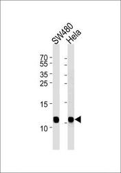
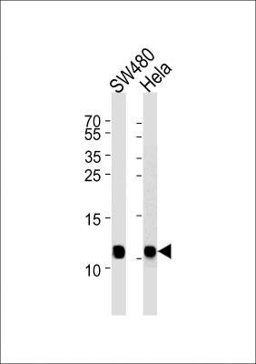
- Western Blot: NDUFA4 Antibody [NBP2-32318] - Western blot analysis in SW480, Hela cell line lysates (35ug/lane). This demonstrates the NDUA4 antibody detected the NDUA4 protein (arrow).
Supportive validation
- Submitted by
- Novus Biologicals (provider)
- Main image
- Experimental details
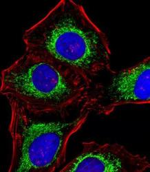
- Immunocytochemistry/Immunofluorescence: NDUFA4 Antibody [NBP2-32318] - Fluorescent image of Hela cells stained with NDUA4 Antibody. It was diluted at 1:25 dilution. An Alexa Fluor 488-conjugated goat anti-rabbit lgG at 1:400 dilution was used as the secondary antibody (green). DAPI was used to stain the cell nuclear (blue). Cytoplasmic actin was counterstained with Alexa Fluor 555 conjugated with Phalloidin (red).

Supportive validation
- Submitted by
- Novus Biologicals (provider)
- Main image

- Experimental details
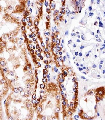
- Immunohistochemistry: NDUFA4 Antibody [NBP2-32318] - Immunohistochemical analysis of paraffin-embedded H. kidney section using NDUA4 Antibody. It was diluted at 1:25 dilution. An undiluted biotinylated goat polyvalent antibody was used as the secondary, followed by DAB staining.