Explore
 Validate
 Learn
Learn Immunohistochemistry
Antibody data
- Antibody Data
- Antigen structure
- References [4]
- Comments [0]
- Validations
- Immunohistochemistry [4]
- Other assay [8]
Submit
Validation data
Reference
Comment
Report error
- Product number
- PA5-60958 - Provider product page

- Provider
- Invitrogen Antibodies
- Product name
- COL6A6 Polyclonal Antibody
- Antibody type
- Polyclonal
- Antigen
- Recombinant protein fragment
- Description
- Immunogen sequence: ALREVEHYFR PDMGSRINTG TPQVLLVLTD GQSQDEVAQA AEALRHRGID IYSVGIGDVD DQQLIQITGT AEKKLTVHNF DELKKVNKRI VRN Highest antigen sequence identity to the following orthologs: Mouse - 87%, Rat - 90%.
- Reactivity
- Human
- Host
- Rabbit
- Isotype
- IgG
- Vial size
- 100 μL
- Concentration
- 0.1 mg/mL
- Storage
- Store at 4°C short term. For long term storage, store at -20°C, avoiding freeze/thaw cycles.
Submitted references Establishment and validation of a novel invasion-related gene signature for predicting the prognosis of ovarian cancer.
COL6A6 inhibits the proliferation and metastasis of non-small cell lung cancer through the JAK signalling pathway.
Cancer-Associated Fibroblast Subgroups Showing Differential Promoting Effect on HNSCC Progression.
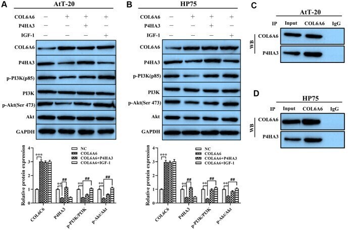
COL6A6 interacted with P4HA3 to suppress the growth and metastasis of pituitary adenoma via blocking PI3K-Akt pathway.
Liang L, Li J, Yu J, Liu J, Xiu L, Zeng J, Wang T, Li N, Wu L
Cancer cell international 2022 Mar 15;22(1):118
Cancer cell international 2022 Mar 15;22(1):118
COL6A6 inhibits the proliferation and metastasis of non-small cell lung cancer through the JAK signalling pathway.
Qiao H, Feng Y, Tang H
Translational cancer research 2021 Oct;10(10):4514-4522
Translational cancer research 2021 Oct;10(10):4514-4522
Cancer-Associated Fibroblast Subgroups Showing Differential Promoting Effect on HNSCC Progression.
Kang SH, Oh SY, Lee HJ, Kwon TG, Kim JW, Lee ST, Choi SY, Hong SH
Cancers 2021 Feb 6;13(4)
Cancers 2021 Feb 6;13(4)
COL6A6 interacted with P4HA3 to suppress the growth and metastasis of pituitary adenoma via blocking PI3K-Akt pathway.
Long R, Liu Z, Li J, Yu H
Aging 2019 Oct 17;11(20):8845-8859
Aging 2019 Oct 17;11(20):8845-8859
No comments: Submit comment
Supportive validation
- Submitted by
- Invitrogen Antibodies (provider)
- Main image
- Experimental details
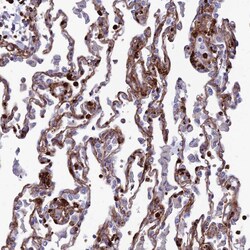
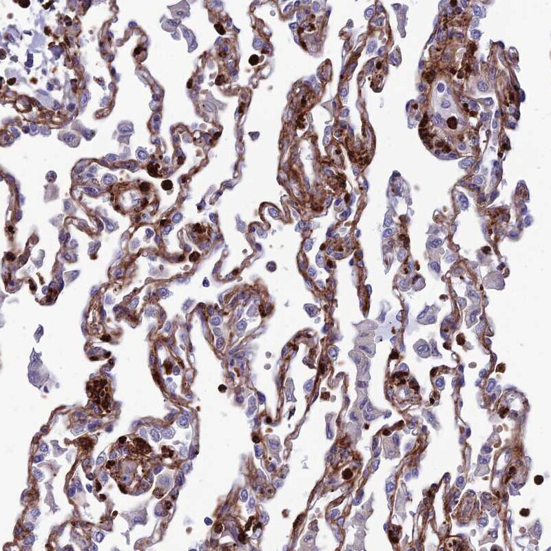
- Immunohistochemical staining of COL6A6 in human lung using COL6A6 Polyclonal Antibody (Product # PA5-60958) shows moderate to strong membranous positivity in pneumocytes.
- Submitted by
- Invitrogen Antibodies (provider)
- Main image
- Experimental details
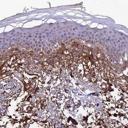
- Immunohistochemical staining of COL6A6 in human skin using COL6A6 Polyclonal Antibody (Product # PA5-60958) shows moderate positivity in extracellular matrix.

- Submitted by
- Invitrogen Antibodies (provider)
- Main image
- Experimental details
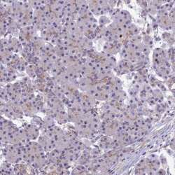
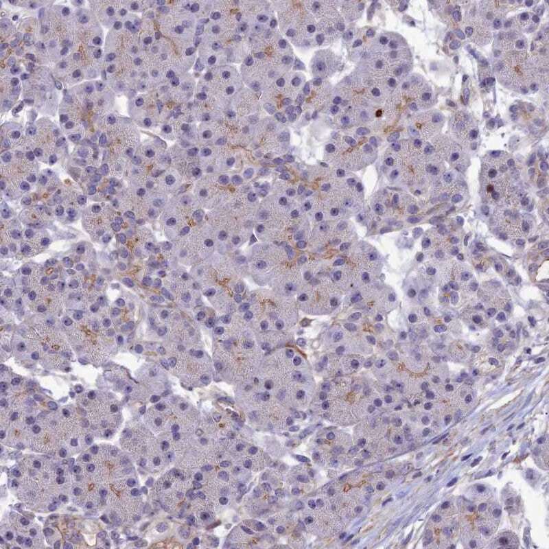
- Immunohistochemical staining of COL6A6 in human pancreas using COL6A6 Polyclonal Antibody (Product # PA5-60958) shows very weak positivity in exocrine glandular cells.
- Submitted by
- Invitrogen Antibodies (provider)
- Main image
- Experimental details
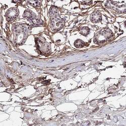
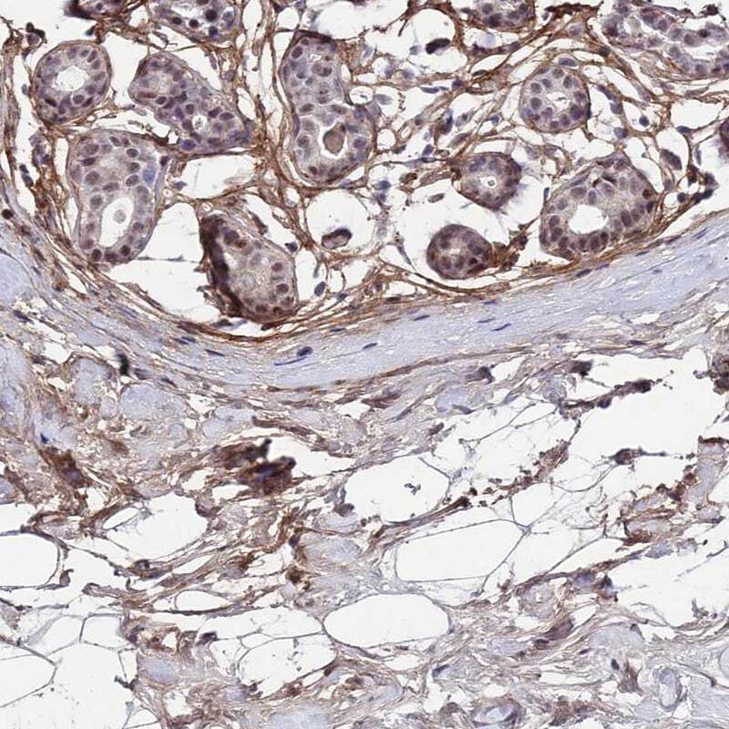
- Immunohistochemical staining of COL6A6 in human breast using COL6A6 Polyclonal Antibody (Product # PA5-60958) shows moderate positivity in extracellular matrix.
Supportive validation
- Submitted by
- Invitrogen Antibodies (provider)
- Main image
- Experimental details
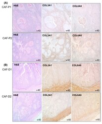
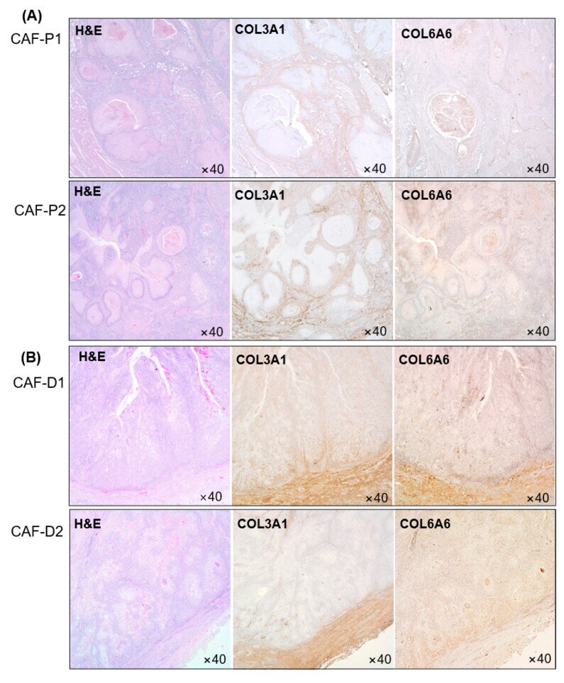
- Figure 5 Collagen protein expression in HNSCC patients' tissues. COL3A1 and COL6A6 protein expression was evaluated in CAF-P ( A ) and CAF-D ( B ) tissues from patients with HNSCC.
- Submitted by
- Invitrogen Antibodies (provider)
- Main image
- Experimental details
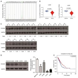
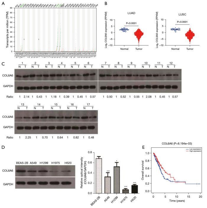
- Figure 1 The expression of COL6A6 in NSCLC tissues. (A) COL6A6 is differentially expressed in tumors compared to normal tissues according to the GEPIA database. (B) The expression of COL6A6 in LUAD and LUSC in TCGA database (P
- Submitted by
- Invitrogen Antibodies (provider)
- Main image
- Experimental details
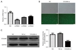
- Figure 2 Verification of the transfection efficiency of COL6A6 knockdown in NSCLC cells. (A) Lentiviral siRNA targeting COL6A6 was transfected into H1299 cells. (B) More than 80% of the cells were green fluorescent protein (GFP) positive and were transfected successfully (x100). Scale bar: 100 um. (C) Western blotting assays were used to detect the expression of COL6A6 in H1299 cells transfected with siRNA. NC: uninfected H1299 cells; NC-si: negative control siRNA transfected H1299 cells; COL6A6-si: COL6A6 siRNA transfected H1299 cells. Data are expressed as mean +- SE. **, P

- Submitted by
- Invitrogen Antibodies (provider)
- Main image
- Experimental details
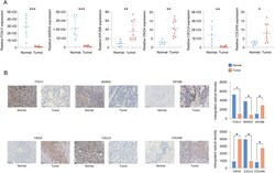
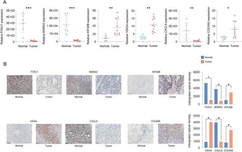
- The qPCR ( A ) and IHC ( B ) results showed that FOXJ1, MXRA5 and CXCL9 expression was low and that KIF26B, VSIG4 and COL6A6 expression was high in OC tissues
- Submitted by
- Invitrogen Antibodies (provider)
- Main image
- Experimental details
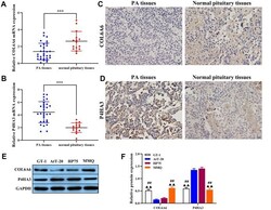
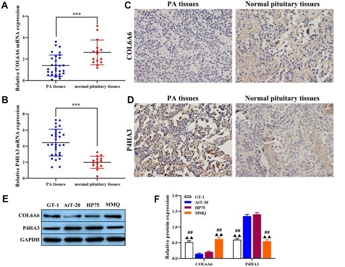
- Figure 2 The expression of COL6A6 and P4HA3 in PA tissues and cell lines. ( A and B ) RT-qPCR was used to detect the expression of COL6A6 and P4HA3 in PA tissues and normal pituitary tissues; ( C and D ) Immunohistochemistry staining was applied to detect the expression of COL6A6 and P4HA3 in PA tissues and normal pituitary tissues; ( E and F ) Western blotting was used to evaluate the expression of COL6A6 and P4HA3 in PA cell lines. *** P
- Submitted by
- Invitrogen Antibodies (provider)
- Main image
- Experimental details
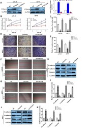
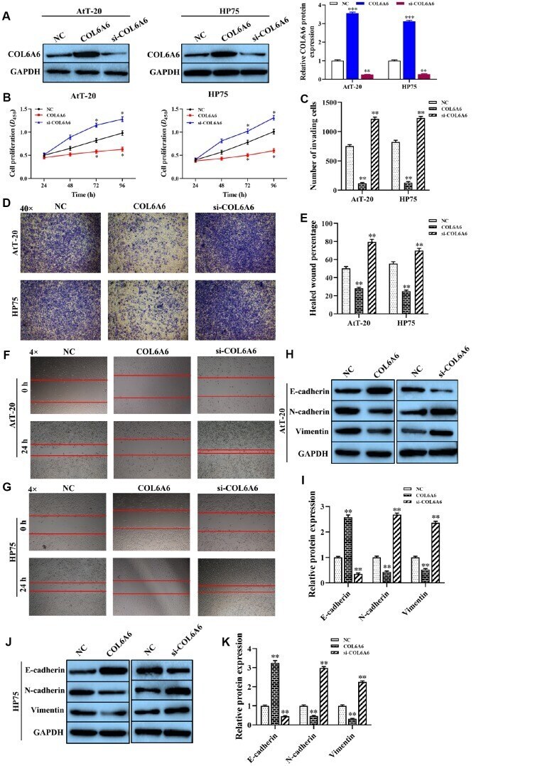
- Figure 3 The effect of COL6A6 on the proliferation, invasion, migration, and EMT of AtT-20 and HP75 cells . ( A ) The expression of COL6A6 protein was determined by western blotting; ( B ) The proliferation ability of AtT-20 and HP75 cells were measured by CCK-8 assay; ( C and D ) The invasion of AtT-20 and HP75 cells were measured by Transwell assay (40x); ( E - G ) The migration ability of AtT-20 and HP75 cells were evaluated by wound healing assay (4x); ( H - K ) The expression of EMT related proteins in AtT-20 and HP75 cells were detected by western blotting. * P
- Submitted by
- Invitrogen Antibodies (provider)
- Main image
- Experimental details
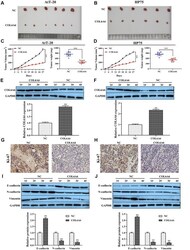
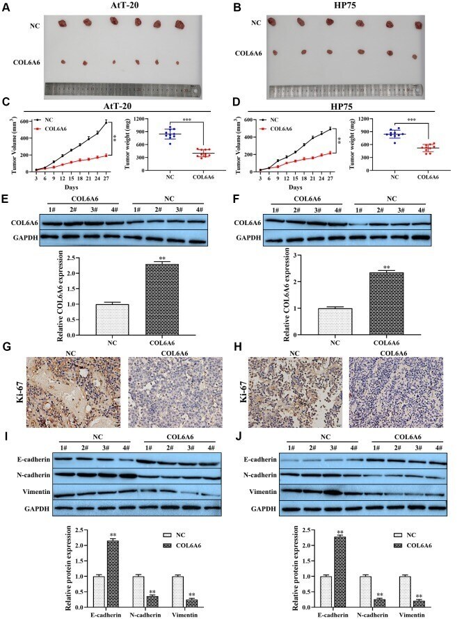
- Figure 4 The effect of COL6A6 on the growth and metastasis of xenograft tumor . ( A and B ) The tumor size was obtained from nude mice; ( C and D ) Tumor volume and tumor weight of nude mice were analyzed; ( E and F ) The expression of COL6A6 in xenograft tumor tissues were detected by western blotting; ( G and H ) The expression of Ki-67 in xenograft tumor tissues were detected by immunohistochemistry staining; ( I and J ) The expression of E-cadherin, vimentin, and N-cadherin were measured by western blotting. ** P
- Submitted by
- Invitrogen Antibodies (provider)
- Main image

- Experimental details
- Figure 5 Effect of COL6A6 on the PI3K-Akt signaling pathway through interacting with P4HA3 . ( A and B ) Western blotting was used to detect the expression of COL6A6, P4HA3, PI3K-Akt pathway-related protein in AtT-20 and HP75 cells; ( C and D ) Coimmunoprecipitation to validate the COL6A6-P4HA3 interaction. ** P