Explore
 Validate
 Learn
Learn Immunocytochemistry
 Immunohistochemistry
Antibody data
- Antibody Data
- Antigen structure
- References [1]
- Comments [0]
- Validations
- Immunohistochemistry [1]
Submit
Validation data
Reference
Comment
Report error
- Product number
- MAB6088 - Provider product page

- Provider
- R&D Systems
- Product name
- Human Neuroligin 3/NLGN3 Antibody
- Antibody type
- Monoclonal
- Description
- Protein A or G purified from hybridoma culture supernatant. Detects human Neuroligin 3 in direct ELISAs. In this format, 100% cross-reactivity with recombinant human (rh) Neuroligin 3 variant 2 and recombinant rat Neuroligin 3 variant 2 and no cross-reactivity with rhNeuroligin 1 variant 2, rhNeuroligin 2, rhNeuroligin 4, recombinant rat Neuroligin 1, 1 variant 2, 2 variant 2, or 3 variant 4 is observed.
- Reactivity
- Human
- Host
- Mouse
- Conjugate
- Unconjugated
- Antigen sequence
NP_061850- Isotype
- IgG
- Antibody clone number
- 566209
- Vial size
- 100 ug
- Concentration
- LYOPH
- Storage
- Use a manual defrost freezer and avoid repeated freeze-thaw cycles. 12 months from date of receipt, -20 to -70 °C as supplied. 1 month, 2 to 8 °C under sterile conditions after reconstitution. 6 months, -20 to -70 °C under sterile conditions after reconstitution.
Submitted references Global Analysis of Protein Expression of Inner Ear Hair Cells.
Hickox AE, Wong AC, Pak K, Strojny C, Ramirez M, Yates JR 3rd, Ryan AF, Savas JN
The Journal of neuroscience : the official journal of the Society for Neuroscience 2017 Feb 1;37(5):1320-1339
The Journal of neuroscience : the official journal of the Society for Neuroscience 2017 Feb 1;37(5):1320-1339
No comments: Submit comment
Supportive validation
- Submitted by
- R&D Systems (provider)
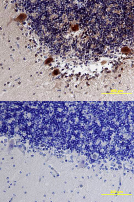
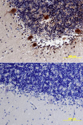
- Main image
- Experimental details
- Neuroligin 3/NLGN3 in Human Brain. Neuroligin 3/NLGN3 was detected in immersion fixed paraffin-embedded sections of human brain (cerebellum) using Human Neuroligin 3/NLGN3 Monoclonal Antibody (Catalog # MAB6088) at 15 µg/mL overnight at 4 °C. Tissue was stained using the Anti-Mouse HRP-DAB Cell & Tissue Staining Kit (brown; Catalog # CTS002) and counterstained with hematoxylin (blue). Lower panel shows a lack of labeling if primary antibodies are omitted and tissue is stained only with secondary antibody followed by incubation with detection reagents. View our protocol for Chromogenic IHC Staining of Paraffin-embedded Tissue Sections.