 Explore
 Validate
 Learn
Learn Western blot
 Immunocytochemistry
Antibody data
- Antibody Data
- Antigen structure
- References [5]
- Comments [0]
- Validations
- Immunocytochemistry [1]
- Immunohistochemistry [1]
- Blocking/Neutralizing [1]
Submit
Validation data
Reference
Comment
Report error
- Product number
- MAB301-100 - Provider product page

- Provider
- R&D Systems
- Product name
- Human IL-3R alpha/CD123 Antibody
- Antibody type
- Monoclonal
- Description
- Protein A or G purified from hybridoma culture supernatant. Detects human IL-3 R alpha/CD123 in direct ELISAs and Western blots.
- Reactivity
- Human
- Host
- Mouse
- Conjugate
- Unconjugated
- Antigen sequence
P26951- Isotype
- IgG
- Antibody clone number
- 32703
- Vial size
- 100 ug
- Storage
- Use a manual defrost freezer and avoid repeated freeze-thaw cycles. 12 months from date of receipt, -20 to -70 °C as supplied. 1 month, 2 to 8 °C under sterile conditions after reconstitution. 6 months, -20 to -70 °C under sterile conditions after reconstitution.
Submitted references A dual role for the N-terminal domain of the IL-3 receptor in cell signalling.
Interleukin-3 enhances the migration of human mesenchymal stem cells by regulating expression of CXCR4.
Human Basophils Modulate Plasma Cell Differentiation and Maturation.
Graves' disease is associated with a defective expression of the immune regulatory molecule galectin-9 in antigen-presenting dendritic cells.
Human T-cell leukemia virus type 2 induces survival and proliferation of CD34(+) TF-1 cells through activation of STAT1 and STAT5 by secretion of interferon-gamma and granulocyte macrophage-colony-stimulating factor.
Broughton SE, Hercus TR, Nero TL, Kan WL, Barry EF, Dottore M, Cheung Tung Shing KS, Morton CJ, Dhagat U, Hardy MP, Wilson NJ, Downton MT, Schieber C, Hughes TP, Lopez AF, Parker MW
Nature communications 2018 Jan 26;9(1):386
Nature communications 2018 Jan 26;9(1):386
Interleukin-3 enhances the migration of human mesenchymal stem cells by regulating expression of CXCR4.
Barhanpurkar-Naik A, Mhaske ST, Pote ST, Singh K, Wani MR
Stem cell research & therapy 2017 Jul 14;8(1):168
Stem cell research & therapy 2017 Jul 14;8(1):168
Human Basophils Modulate Plasma Cell Differentiation and Maturation.
Dijkstra D, Meyer-Bahlburg A
Journal of immunology (Baltimore, Md. : 1950) 2017 Jan 1;198(1):229-238
Journal of immunology (Baltimore, Md. : 1950) 2017 Jan 1;198(1):229-238
Graves' disease is associated with a defective expression of the immune regulatory molecule galectin-9 in antigen-presenting dendritic cells.
Leskela S, Serrano A, de la Fuente H, Rodríguez-Muñoz A, Ramos-Levi A, Sampedro-Nuñez M, Sánchez-Madrid F, González-Amaro R, Marazuela M
PloS one 2015;10(4):e0123938
PloS one 2015;10(4):e0123938
Human T-cell leukemia virus type 2 induces survival and proliferation of CD34(+) TF-1 cells through activation of STAT1 and STAT5 by secretion of interferon-gamma and granulocyte macrophage-colony-stimulating factor.
Bovolenta C, Pilotti E, Mauri M, Turci M, Ciancianaini P, Fisicaro P, Bertazzoni U, Poli G, Casoli C
Blood 2002 Jan 1;99(1):224-31
Blood 2002 Jan 1;99(1):224-31
No comments: Submit comment
Supportive validation
- Submitted by
- R&D Systems (provider)
- Main image

- Experimental details
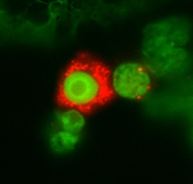
- IL-3 R alpha/CD123 in Human Peripheral Blood Lymphocytes. IL-3 R alpha/CD123 was detect-ed in immersion fixed human peripheral blood lymphocytes using 2 µg/mL Mouse Anti-Human IL-3 R alpha/CD123 Monoclonal Antibody (Catalog # MAB301) for 3 hours at room temperature. Cells were stained (red) and counter-stained (green). View our protocol for Fluorescent ICC Staining of Non-adherent Cells.
Supportive validation
- Submitted by
- R&D Systems (provider)
- Main image

- Experimental details
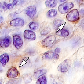
- IL-3 R alpha/CD123 in Human Tonsil. IL-3 R alpha/CD123 was detected in immersion fixed paraffin-embedded sections of human tonsil using 15 µg/mL Mouse Anti-Human IL-3 R alpha/CD123 Monoclonal Antibody (Catalog # MAB301) overnight at 4 °C. Tissue was stained with the Anti-Mouse HRP-DAB Cell & Tissue Staining Kit (brown; Catalog # CTS002) and counter-stained with hematoxylin (blue). View our protocol for Chromogenic IHC Staining of Paraffin-embedded Tissue Sections.
Supportive validation
- Submitted by
- R&D Systems (provider)
- Main image
- Experimental details
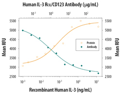
- Cell Proliferation Induced by IL-3 and Neutralization by Human IL-3 R alpha/CD123 Antibody. Recombinant Human IL-3 (Catalog # 203-IL) stimulates proliferation in the TF-1 human erythroleukemic cell line in a dose-dependent manner (orange line). Proliferation elicited by Recombinant Human IL-3 (0.5 ng/mL) is neutralized (green line) by increasing concentrations of Mouse Anti-Human IL-3 R alpha/CD123 Monoclonal Antibody (Catalog # MAB301). The ND50 is typically 0.6-1.2 µg/mL.
