 Explore
 Validate
 Learn
Learn Flow cytometry
Antibody data
- Antibody Data
- Antigen structure
- References [6]
- Comments [0]
- Validations
- Flow cytometry [1]
- Other assay [1]
Submit
Validation data
Reference
Comment
Report error
- Product number
- 48-1238-42 - Provider product page

- Provider
- Invitrogen Antibodies
- Product name
- CD123 Monoclonal Antibody (7G3), eFluor™ 450, eBioscience™
- Antibody type
- Monoclonal
- Antigen
- Other
- Description
- Description: This 7G3 monoclonal antibody reacts with human and non-human primate CD123, the alpha chain of the IL-3 receptor. This 60-70 kDa transmembrane protein binds to IL-3 with low affinity by itself, and when associated with CD131 (the common beta chain) binds IL-3 with high affinity. CD123 is expressed by myeloid precursors, macrophages, dendritic cells, mast cells, basophils, and megakaryocytes. In addition, this molecule can be detected on CD34+CD38- hematopoietic stem cells and acute myeloid leukemic (AML) cells. The 7G3 antibody has been demonstrated to inhibit IL-3-mediated cell proliferation and homing of AML leukemic stem cells. Crossblocking studies suggest that 7G3 recognizes a different epitope from 6H6. The 7G3 antibody has been reported to crossreact to chimpanzee, cynomologous monkey, olive baboon, pigtailed macaque, rhesus macaque, and sooty mangabey. Applications Reported: This 7G3 antibody has been reported for use in flow cytometric analysis. Applications Tested: This 7G3 antibody has been pre-titrated and tested by flow cytometric analysis of normal human peripheral blood cells. This can be used at 5 µL (0.5 µg) per test. A test is defined as the amount (µg) of antibody that will stain a cell sample in a final volume of 100 µL. Cell number should be determined empirically but can range from 10^5 to 10^8 cells/test. eFluor® 450 is an alternative to Pacific Blue®. eFluor® 450 emits at 445 nm and is excited with the Violet laser (405 nm). Please make sure that your instrument is capable of detecting this fluorochome. Excitation: 405 nm; Emission: 445 nm; Laser: Violet Laser. Filtration: 0.2 µm post-manufacturing filtered.
- Reactivity
- Human
- Host
- Mouse
- Isotype
- IgG
- Antibody clone number
- 7G3
- Vial size
- 100 Tests
- Concentration
- 5 μL/Test
- Storage
- 4°C, store in dark, DO NOT FREEZE!
Submitted references Improving hematopoietic recovery through modeling and modulation of the mesenchymal stromal cell secretome.
Expression of dendritic cell markers CD11c/BDCA-1 and CD123/BDCA-2 in coronary artery disease upon activation in whole blood.
Monoclonal antibody-mediated targeting of CD123, IL-3 receptor alpha chain, eliminates human acute myeloid leukemic stem cells.
Surface phenotype and rapid quantification of blood dendritic cell subsets in the rhesus macaque.
Parallel loss of myeloid and plasmacytoid dendritic cells from blood and lymphoid tissue in simian AIDS.
A flow cytometric immune function assay for human peripheral blood dendritic cells.
Liu FD, Tam K, Pishesha N, Poon Z, Van Vliet KJ
Stem cell research & therapy 2018 Oct 24;9(1):268
Stem cell research & therapy 2018 Oct 24;9(1):268
Expression of dendritic cell markers CD11c/BDCA-1 and CD123/BDCA-2 in coronary artery disease upon activation in whole blood.
Van Brussel I, Van Vré EA, De Meyer GR, Vrints CJ, Bosmans JM, Bult H
Journal of immunological methods 2010 Oct 31;362(1-2):168-75
Journal of immunological methods 2010 Oct 31;362(1-2):168-75
Monoclonal antibody-mediated targeting of CD123, IL-3 receptor alpha chain, eliminates human acute myeloid leukemic stem cells.
Jin L, Lee EM, Ramshaw HS, Busfield SJ, Peoppl AG, Wilkinson L, Guthridge MA, Thomas D, Barry EF, Boyd A, Gearing DP, Vairo G, Lopez AF, Dick JE, Lock RB
Cell stem cell 2009 Jul 2;5(1):31-42
Cell stem cell 2009 Jul 2;5(1):31-42
Surface phenotype and rapid quantification of blood dendritic cell subsets in the rhesus macaque.
Brown KN, Barratt-Boyes SM
Journal of medical primatology 2009 Aug;38(4):272-8
Journal of medical primatology 2009 Aug;38(4):272-8
Parallel loss of myeloid and plasmacytoid dendritic cells from blood and lymphoid tissue in simian AIDS.
Brown KN, Trichel A, Barratt-Boyes SM
Journal of immunology (Baltimore, Md. : 1950) 2007 Jun 1;178(11):6958-67
Journal of immunology (Baltimore, Md. : 1950) 2007 Jun 1;178(11):6958-67
A flow cytometric immune function assay for human peripheral blood dendritic cells.
Willmann K, Dunne JF
Journal of leukocyte biology 2000 Apr;67(4):536-44
Journal of leukocyte biology 2000 Apr;67(4):536-44
No comments: Submit comment
Supportive validation
- Submitted by
- Invitrogen Antibodies (provider)
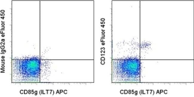
- Main image
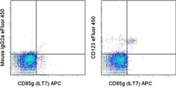
- Experimental details
- Staining of normal human peripheral blood cells with Anti-Human CD85g (ILT7) APC (Product # 17-5179-42) and Mouse IgG2a K Isotype Control eFluor® 450 (Product # 48-4724-82) (left) or Anti-Human/Non-Human Primate CD123 eFluor® 450 (right). Total viable cells were used for analysis.
Supportive validation
- Submitted by
- Invitrogen Antibodies (provider)
- Main image
- Experimental details
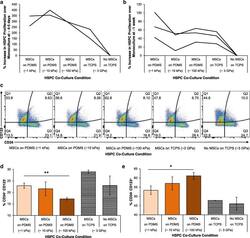
- Fig. 3 In vitro hematopoietic recovery: HSPC and MSC co-culture. Hematopoietic stem and progenitors (HSPCs) were grown in direct contact co-culture with MSCs grown on 1, 10, 100 kPa PDMS and TCPS. Proliferation of HSPCs in these co-culture conditions relative to monoculture were determined at ( a ) 4-5 days and ( b ) 1 week after co-culture. Individual lines represent replicate experiments. c HSPC surface marker expression of CD123 and CD34 assayed using flow cytometry. d % expression of CD34 + and CD123 - cells as mean +- standard deviations. e % expression of CD34 - and CD123 + cells as mean +- standard deviations. d - e Co-culture on PDMS of increasing stiffness (1, 10, 100 kPa) indicated in darkening shades of orange , respectively. Co-culture and monoculture of HSPCs on tissue culture polystyrene are shown as with black- and white-striped bars as distinct current standard protocols for comparison. Significant differences were calculated with one-tailed Student's t tests with unequal variance, (* p < 0.01, ** p < 0.005). Data are plotted as mean +- standard deviation, N = 3 across replicate wells
