Explore
 Validate
 Learn
Learn Western blot
 Immunocytochemistry
Antibody data
- Antibody Data
- Antigen structure
- References [3]
- Comments [0]
- Validations
- Immunocytochemistry [2]
- Immunohistochemistry [1]
- Other assay [1]
Submit
Validation data
Reference
Comment
Report error
- Product number
- PA5-28901 - Provider product page

- Provider
- Invitrogen Antibodies
- Product name
- MINK1 Polyclonal Antibody
- Antibody type
- Polyclonal
- Antigen
- Recombinant full-length protein
- Description
- Recommended positive controls: HepG2. Predicted reactivity: Mouse (97%), Rat (97%), Zebrafish (80%), Xenopus laevis (85%), Bovine (98%). Store product as a concentrated solution. Centrifuge briefly prior to opening the vial.
- Reactivity
- Human
- Host
- Rabbit
- Isotype
- IgG
- Vial size
- 100 μL
- Concentration
- 1 mg/mL
- Storage
- Store at 4°C short term. For long term storage, store at -20°C, avoiding freeze/thaw cycles.
Submitted references Prickle promotes the formation and maintenance of glutamatergic synapses by stabilizing the intercellular planar cell polarity complex.
Identification of Endogenous Adenomatous Polyposis Coli Interaction Partners and β-Catenin-Independent Targets by Proteomics.
The homophilic receptor PTPRK selectively dephosphorylates multiple junctional regulators to promote cell-cell adhesion.
Ban Y, Yu T, Feng B, Lorenz C, Wang X, Baker C, Zou Y
Science advances 2021 Oct 8;7(41):eabh2974
Science advances 2021 Oct 8;7(41):eabh2974
Identification of Endogenous Adenomatous Polyposis Coli Interaction Partners and β-Catenin-Independent Targets by Proteomics.
Popow O, Paulo JA, Tatham MH, Volk MS, Rojas-Fernandez A, Loyer N, Newton IP, Januschke J, Haigis KM, Näthke I
Molecular cancer research : MCR 2019 Sep;17(9):1828-1841
Molecular cancer research : MCR 2019 Sep;17(9):1828-1841
The homophilic receptor PTPRK selectively dephosphorylates multiple junctional regulators to promote cell-cell adhesion.
Fearnley GW, Young KA, Edgar JR, Antrobus R, Hay IM, Liang WC, Martinez-Martin N, Lin W, Deane JE, Sharpe HJ
eLife 2019 Mar 29;8
eLife 2019 Mar 29;8
No comments: Submit comment
Supportive validation
- Submitted by
- Invitrogen Antibodies (provider)
- Main image

- Experimental details
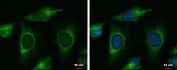
- MINK1 Polyclonal Antibody detects MAP4K6 protein at cytoplasm by immunofluorescent analysis. Sample: HeLa cells were fixed in ice-cold MeOH for 5 min. Green: MAP4K6 protein stained by MINK1 Polyclonal Antibody (Product # PA5-28901) diluted at 1:500. Blue: Hoechst 33342 staining.

- Submitted by
- Invitrogen Antibodies (provider)
- Main image
- Experimental details
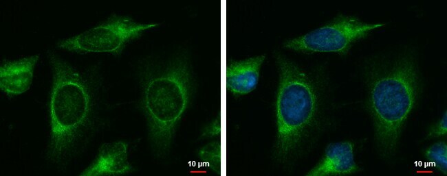
- MINK1 Polyclonal Antibody detects MAP4K6 protein at cytoplasm by immunofluorescent analysis. Sample: HeLa cells were fixed in ice-cold MeOH for 5 min. Green: MAP4K6 protein stained by MINK1 Polyclonal Antibody (Product # PA5-28901) diluted at 1:500. Blue: Hoechst 33342 staining.
Supportive validation
- Submitted by
- Invitrogen Antibodies (provider)
- Main image
- Experimental details
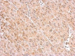
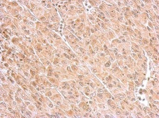
- MINK1 Polyclonal Antibody detects MINK1 protein at cytosol on U87 xenograft by immunohistochemical analysis. Sample: Paraffin-embedded U87 xenograft. MINK1 Polyclonal Antibody (Product # PA5-28901) dilution: 1:500. Antigen Retrieval: EDTA based buffer, pH 8.0, 15 min.
Supportive validation
- Submitted by
- Invitrogen Antibodies (provider)
- Main image
- Experimental details
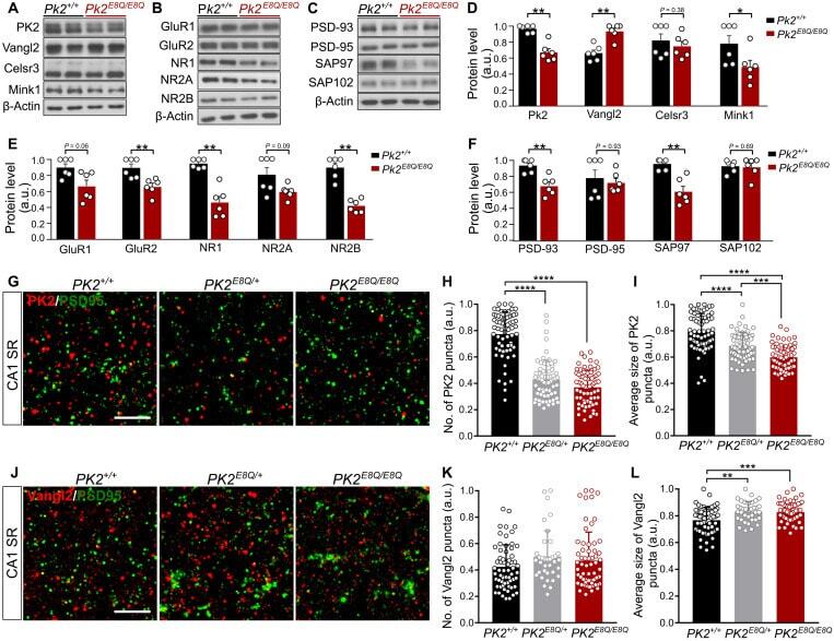
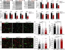
- Fig. 4. Altered composition of synaptic proteins in the PK2 E8Q/E8Q mice. ( A to C ) Western blot analysis of PCP components, regulator of PCP signaling, Mink1, alpha-amino-3-hydroxy-5-methyl-4-isoxazole propionic acid receptor (AMPAR)/N-methyl-D-aspartate receptor (NMDAR) subunits, and MAGUK family proteins in the crude synaptosomes of cortex and HPC from P14 PK2 E8Q/E8Q mice and WT littermates. ( D to F ) Quantitation of protein levels shown in (A) to (C). Each data point represents one animal. Data were normalized by dividing each value to the maximum value in each independent experiment. PK2 +/+ , n = 6; PK2 E8Q/E8Q , n = 6, Mann-Whitney U test, * P < 0.05 and ** P < 0.01. Error bars represent SEM. ( G to L ) Immunofluorescence staining of PK2 and Vangl2 in CA1 SR of P14 mice. To quantify the number (H) and the average size (I) of PK2 puncta, 66 image stacks of CA1 SR were taken from three PK2 +/+ animals, 66 image stacks from three PK2 E8Q/+ animals, and 68 image stacks from three PK2 E8Q/E8Q animals. To quantify the number (K) and the average size (L) of Vangl2 puncta, 62 image stacks were taken from three PK2 +/+ animals, 36 image stacks from two PK2 E8Q/+ animals, and 63 image stacks from PK2 E8Q/E8Q animals. Data were normalized by dividing each value to the maximum value in each independent experiment. Scale bars, 5 mum. Kruskal-Wallis test was used to compare the means of three groups, and Dunn's test was used to perform multiple comparisons. ** P < 0.01, *** P < 0