Explore
 Validate
 Learn
Learn37-7700
antibody from Invitrogen Antibodies
Targeting: UBQLN2
Chap1, CHAP1/DSK2, Dsk2, LIC-2, N4BP4, PLIC-2, PLIC2, RIHFB2157
 Western blot
 ELISA
ELISA Immunocytochemistry Immunoprecipitation Immunohistochemistry Flow cytometry Other assay
Antibody data
- Antibody Data
- Antigen structure
- References [7]
- Comments [0]
- Validations
- Immunocytochemistry [2]
- Immunohistochemistry [3]
- Flow cytometry [1]
- Other assay [6]
Submit
Validation data
Reference
Comment
Report error
- Product number
- 37-7700 - Provider product page

- Provider
- Invitrogen Antibodies
- Product name
- Ubiquilin 2 Monoclonal Antibody (3D5E2)
- Antibody type
- Monoclonal
- Antigen
- Recombinant full-length protein
- Reactivity
- Human, Mouse
- Host
- Mouse
- Isotype
- IgG
- Antibody clone number
- 3D5E2
- Vial size
- 100 μg
- Concentration
- 0.5 mg/mL
- Storage
- -20°C
Submitted references Human Ubiquilin 2 and TDP-43 copathology drives neurodegeneration in transgenic Caenorhabditis elegans.
Overexpression of UBQLN1 reduces neuropathology in the P497S UBQLN2 mouse model of ALS/FTD.
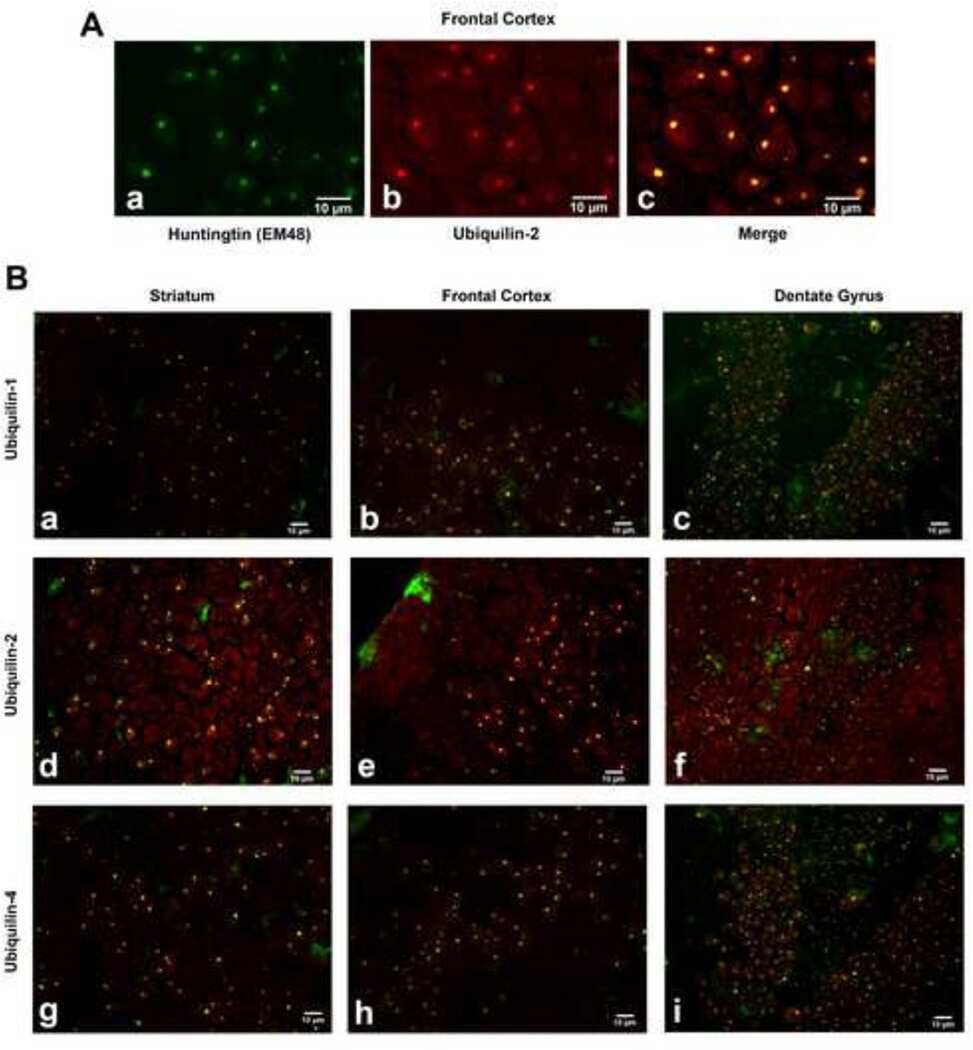
Signature changes in ubiquilin expression in the R6/2 mouse model of Huntington's disease.
Ubiquilin-1 overexpression increases the lifespan and delays accumulation of Huntingtin aggregates in the R6/2 mouse model of Huntington's disease.
Nuclear proteasomes carry a constitutive posttranslational modification which derails SDS-PAGE (but not CTAB-PAGE).
Studies of the role of ubiquitination in the interaction of ubiquilin with the loop and carboxyl terminal regions of presenilin-2.
Ubiquilin 1 modulates amyloid precursor protein trafficking and Abeta secretion.
Saxton AD, Kraemer BC
G3 (Bethesda, Md.) 2021 Aug 7;11(8)
G3 (Bethesda, Md.) 2021 Aug 7;11(8)
Overexpression of UBQLN1 reduces neuropathology in the P497S UBQLN2 mouse model of ALS/FTD.
Wang S, Tatman M, Monteiro MJ
Acta neuropathologica communications 2020 Oct 7;8(1):164
Acta neuropathologica communications 2020 Oct 7;8(1):164
Signature changes in ubiquilin expression in the R6/2 mouse model of Huntington's disease.
Safren N, Chang L, Dziki KM, Monteiro MJ
Brain research 2015 Feb 9;1597:37-46
Brain research 2015 Feb 9;1597:37-46
Ubiquilin-1 overexpression increases the lifespan and delays accumulation of Huntingtin aggregates in the R6/2 mouse model of Huntington's disease.
Safren N, El Ayadi A, Chang L, Terrillion CE, Gould TD, Boehning DF, Monteiro MJ
PloS one 2014;9(1):e87513
PloS one 2014;9(1):e87513
Nuclear proteasomes carry a constitutive posttranslational modification which derails SDS-PAGE (but not CTAB-PAGE).
Pitcher DS, de Mattos-Shipley K, Wang Z, Tzortzis K, Goudevenou K, Flynn H, Bohn G, Rahemtulla A, Roberts I, Snijders AP, Karadimitris A, Kleijnen MF
Biochimica et biophysica acta 2014 Dec;1844(12):2222-8
Biochimica et biophysica acta 2014 Dec;1844(12):2222-8
Studies of the role of ubiquitination in the interaction of ubiquilin with the loop and carboxyl terminal regions of presenilin-2.
Ford DL, Monteiro MJ
Biochemistry 2007 Jul 31;46(30):8827-37
Biochemistry 2007 Jul 31;46(30):8827-37
Ubiquilin 1 modulates amyloid precursor protein trafficking and Abeta secretion.
Hiltunen M, Lu A, Thomas AV, Romano DM, Kim M, Jones PB, Xie Z, Kounnas MZ, Wagner SL, Berezovska O, Hyman BT, Tesco G, Bertram L, Tanzi RE
The Journal of biological chemistry 2006 Oct 27;281(43):32240-53
The Journal of biological chemistry 2006 Oct 27;281(43):32240-53
No comments: Submit comment
Supportive validation
- Submitted by
- Invitrogen Antibodies (provider)
- Main image
- Experimental details
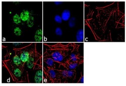
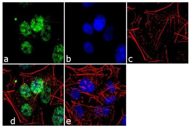
- Immunofluorescence analysis of Ubiquilin / Dsk2 was performed using 70% confluent log phase SH-SY5Y cells. The cells were fixed with 4% paraformaldehyde for 10 minutes, permeabilized with 0.1% Triton™ X-100 for 10 minutes, and blocked with 1% BSA for 1 hour at room temperature. The cells were labeled with Ubiquilin / Dsk2 (3D5E2) Mouse Monoclonal Antibody (Product # 37-7700) at 2µg/mL in 0.1% BSA and incubated for 3 hours at room temperature and then labeled with Goat anti-Mouse IgG (H+L) Superclonal™ Secondary Antibody, Alexa Fluor® 488 conjµgate (Product # A28175) at a dilution of 1:2000 for 45 minutes at room temperature (Panel a: green). Nuclei (Panel b: blue) were stained with SlowFade® Gold Antifade Mountant with DAPI (Product # S36938). F-actin (Panel c: red) was stained with Alexa Fluor® 555 Rhodamine Phalloidin (Product # R415, 1:300). Panel d represents the merged image showing nuclear localization. Panel e shows the no primary antibody control. The images were captured at 60X magnification.

- Submitted by
- Invitrogen Antibodies (provider)
- Main image
- Experimental details
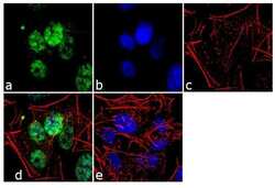
- Immunofluorescence analysis of Ubiquilin / Dsk2 was performed using 70% confluent log phase SH-SY5Y cells. The cells were fixed with 4% paraformaldehyde for 10 minutes, permeabilized with 0.1% Triton™ X-100 for 10 minutes, and blocked with 1% BSA for 1 hour at room temperature. The cells were labeled with Ubiquilin / Dsk2 (3D5E2) Mouse Monoclonal Antibody (Product # 37-7700) at 2µg/mL in 0.1% BSA and incubated for 3 hours at room temperature and then labeled with Goat anti-Mouse IgG (H+L) Superclonal™ Secondary Antibody, Alexa Fluor® 488 conjµgate (Product # A28175) at a dilution of 1:2000 for 45 minutes at room temperature (Panel a: green). Nuclei (Panel b: blue) were stained with SlowFade® Gold Antifade Mountant with DAPI (Product # S36938). F-actin (Panel c: red) was stained with Alexa Fluor® 555 Rhodamine Phalloidin (Product # R415, 1:300). Panel d represents the merged image showing nuclear localization. Panel e shows the no primary antibody control. The images were captured at 60X magnification.
Supportive validation
- Submitted by
- Invitrogen Antibodies (provider)
- Main image

- Experimental details
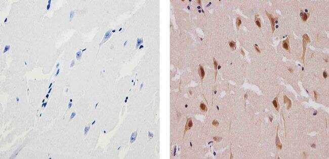
- Immunohistochemistry analysis of Ubiquilin/Dsk2 showing staining in the cytoplasm and nucleus of paraffin-embedded human brain tissue (right) compared to a negative control without primary antibody (left). To expose target proteins, antigen retrieval was performed using 10mM sodium citrate (pH 6.0), microwaved for 8-15 min. Following antigen retrieval, tissues were blocked in 3% H2O2-methanol for 15 min at room temperature, washed with ddH2O and PBS, and then probed with a Ubiquilin/Dsk2 Mouse Monoclonal Antibody (Product # 37-7700) diluted in 3% BSA-PBS at a dilution of 1:100 for 1 hour at 37ºC in a humidified chamber. Tissues were washed extensively in PBST and detection was performed using an HRP-conjugated secondary antibody followed by colorimetric detection using a DAB kit. Tissues were counterstained with hematoxylin and dehydrated with ethanol and xylene to prep for mounting.
- Submitted by
- Invitrogen Antibodies (provider)
- Main image
- Experimental details
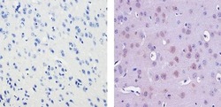
- Immunohistochemistry analysis of Ubiquilin/Dsk2 showing staining in the cytoplasm and nucleus of paraffin-embedded mouse brain tissue (right) compared to a negative control without primary antibody (left). To expose target proteins, antigen retrieval was performed using 10mM sodium citrate (pH 6.0), microwaved for 8-15 min. Following antigen retrieval, tissues were blocked in 3% H2O2-methanol for 15 min at room temperature, washed with ddH2O and PBS, and then probed with a Ubiquilin/Dsk2 Mouse Monoclonal Antibody (Product # 37-7700) diluted in 3% BSA-PBS at a dilution of 1:20 for 1 hour at 37ºC in a humidified chamber. Tissues were washed extensively in PBST and detection was performed using an HRP-conjugated secondary antibody followed by colorimetric detection using a DAB kit. Tissues were counterstained with hematoxylin and dehydrated with ethanol and xylene to prep for mounting.
- Submitted by
- Invitrogen Antibodies (provider)
- Main image
- Experimental details
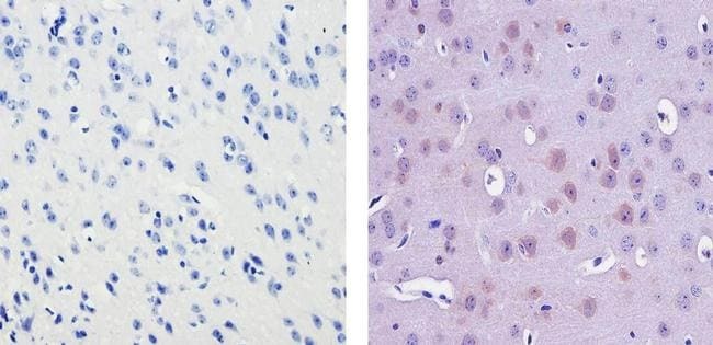
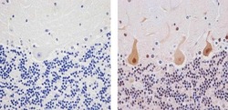
- Immunohistochemistry analysis of Ubiquilin/Dsk2 showing staining in the cytoplasm and nucleus of paraffin-embedded human cerebellum tissue (right) compared to a negative control without primary antibody (left). To expose target proteins, antigen retrieval was performed using 10mM sodium citrate (pH 6.0), microwaved for 8-15 min. Following antigen retrieval, tissues were blocked in 3% H2O2-methanol for 15 min at room temperature, washed with ddH2O and PBS, and then probed with a Ubiquilin/Dsk2 Mouse Monoclonal Antibody (Product # 37-7700) diluted in 3% BSA-PBS at a dilution of 1:20 for 1 hour at 37ºC in a humidified chamber. Tissues were washed extensively in PBST and detection was performed using an HRP-conjugated secondary antibody followed by colorimetric detection using a DAB kit. Tissues were counterstained with hematoxylin and dehydrated with ethanol and xylene to prep for mounting.

Supportive validation
- Submitted by
- Invitrogen Antibodies (provider)
- Main image
- Experimental details
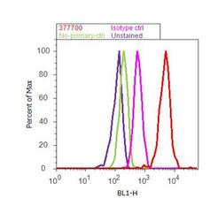
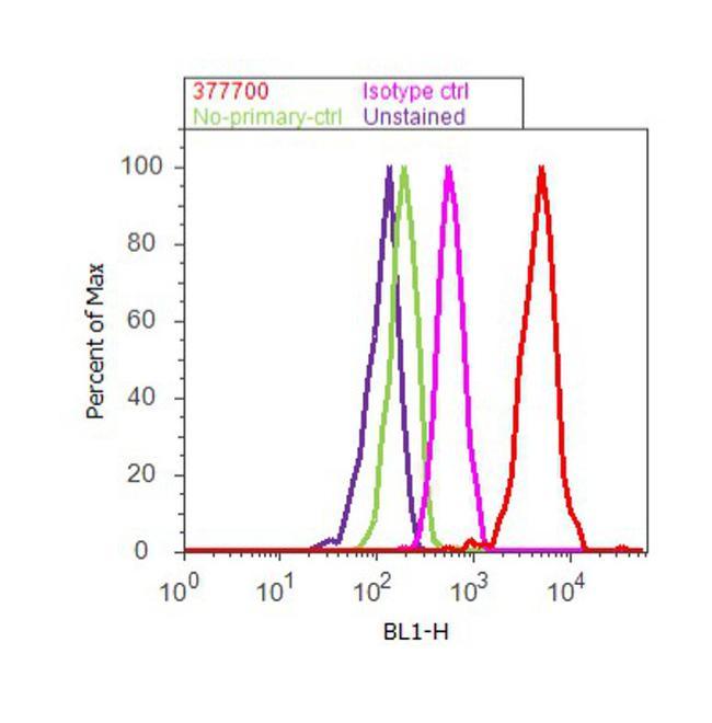
- Flow cytometry analysis of Ubiquilin / Dsk2 was done on SH-SY5Y cells. Cells were fixed with 70% ethanol for 10 minutes, permeabilized with 0.25% Triton™ X-100 for 20 minutes, and blocked with 5% BSA for 30 minutes at room temperature. Cells were labeled with Ubiquilin / Dsk2 Mouse Monoclonal Antibody (Product # 37-7700, red histogram) or with mouse isotype control (pink histogram) at 3-5 µg/million cells in 2.5% BSA. After incubation at room temperature for 2 hours, the cells were labeled with Alexa Fluor® 488 Rabbit Anti-Mouse Secondary Antibody (Product # A11059) at a dilution of 1:400 for 30 minutes at room temperature. The representative 10,000 cells were acquired and analyzed for each sample using an Attune® Acoustic Focusing Cytometer. The purple histogram represents unstained control cells and the green histogram represents no-primary-antibody control..
Supportive validation
- Submitted by
- Invitrogen Antibodies (provider)
- Main image
- Experimental details
- NULL

- Submitted by
- Invitrogen Antibodies (provider)
- Main image
- Experimental details
- NULL
- Submitted by
- Invitrogen Antibodies (provider)
- Main image
- Experimental details
- NULL

- Submitted by
- Invitrogen Antibodies (provider)
- Main image

- Experimental details
- NULL
- Submitted by
- Invitrogen Antibodies (provider)
- Main image

- Experimental details
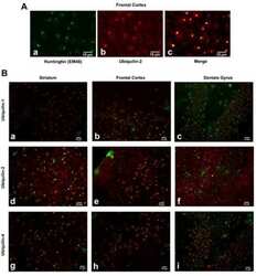
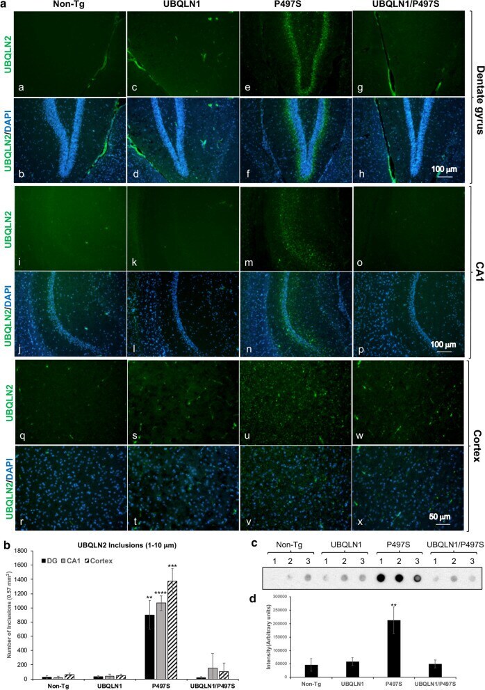
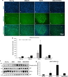
- Fig. 4 UBQLN1 overexpression reduces deposition of UBQLN2 inclusions compared to the brains of P497S mutant mice. a Staining of representative sagittal sections of the dentate gyrus (DG) (a-h), CA1 (i-p) and cortex (q-x) regions of the brain for UBQLN2 and DAPI in the four genotypes at 52 weeks of age. Scale bars shown. b Quantification of the number of UBQLN2 inclusions between 1 and 10 mum in size counted in identical size regions of the DG, CA1 and cortex regions (2 males and 1 female for each genotype). ** P < 0.01, *** P < 0.001, **** P < 0.0001. c Filter-trap assay showing detection of UBQLN2 inclusions in 50 mug of protein brain lysate analyzed for the four genotypes for animals at 52 weeks of age. Samples analyzed were: Non-Tg, 2 male (M) and 1 female (F); UBQLN1 all F; P497S, 1 M, 2 F; UBQLN1/P497S all M. d Quantification of the intensity of UBQLN2 immunoreactivity for the filter shown in c . ** P < 0.01
- Submitted by
- Invitrogen Antibodies (provider)
- Main image
- Experimental details
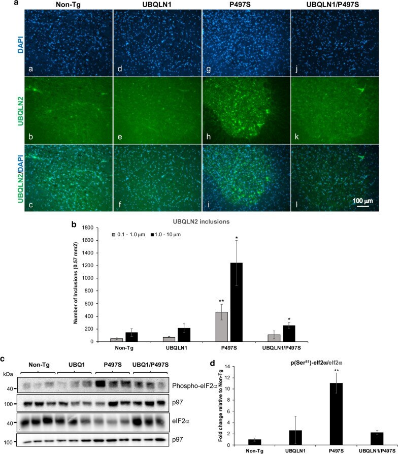
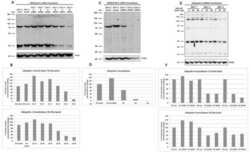
- Fig. 6 Overexpression of UBQLN1 reduces UBQLN2 inclusions and ER stress in the spinal cord. a Immunofluorescence staining of the sections in the ventral horn region of the spinal cord for UBQLN2 and DAPI and of their combined staining. b Quantification of the number of UBQLN2 inclusions based on size in the spinal cord of the four genotypes at 52 weeks of age (2 males and 1 female for each genotype). * P < 0.05, ** P < 0.01. c Immunoblots of SC lysates for the different genotypes probed for the proteins shown. d Quantification of changes in phospho-eIF2alpha after normalizing for expression of total eIF2alpha protein (2 males and 1 female for each genotype). ** P < 0.01