Explore
 Validate
 Learn
Learn Western blot
Antibody data
- Antibody Data
- Antigen structure
- References [1]
- Comments [0]
- Validations
- Western blot [2]
- Immunohistochemistry [1]
- Other assay [1]
Submit
Validation data
Reference
Comment
Report error
- Product number
- PA5-24598 - Provider product page

- Provider
- Invitrogen Antibodies
- Product name
- IDO Polyclonal Antibody
- Antibody type
- Polyclonal
- Antigen
- Synthetic peptide
- Reactivity
- Human, Mouse
- Host
- Rabbit
- Isotype
- IgG
- Vial size
- 400 μL
- Concentration
- 2 mg/mL
- Storage
- Store at 4°C short term. For long term storage, store at -20°C, avoiding freeze/thaw cycles.
Submitted references The extracts of Astragalus membranaceus enhance chemosensitivity and reduce tumor indoleamine 2, 3-dioxygenase expression.
Phacharapiyangkul N, Wu LH, Lee WY, Kuo YH, Wu YJ, Liou HP, Tsai YE, Lee CH
International journal of medical sciences 2019;16(8):1107-1115
International journal of medical sciences 2019;16(8):1107-1115
No comments: Submit comment
Supportive validation
- Submitted by
- Invitrogen Antibodies (provider)
- Main image
- Experimental details
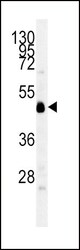
- Western blot analysis of IDO in mouse cerebellum tissue lysates. Samples were incubated with IDO polyclonal antibody (Product # PA5-24598). Lysates: 35 µg/lane. INDO protein (arrow).
- Submitted by
- Invitrogen Antibodies (provider)
- Main image
- Experimental details
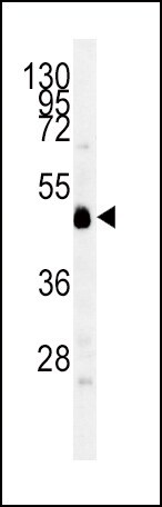
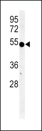
- Western blot analysis of IDO in K562 cell line lysates. Samples were incubated with IDO polyclonal antibody (Product # PA5-24598). Lysates: 35 µg/lane. INDO protein (arrow).
Supportive validation
- Submitted by
- Invitrogen Antibodies (provider)
- Main image

- Experimental details
- Immunohistochemistry analysis of IDO in formalin fixed and paraffin embedded human lung carcinoma. Samples were incubated with IDO polyclonal antibody (Product # PA5-24598) followed by peroxidase conjugation of the secondary antibody and DAB staining. This data demonstrates the use of this antibody for immunohistochemistry. Clinical relevance has not been evaluated.

Supportive validation
- Submitted by
- Invitrogen Antibodies (provider)
- Main image
- Experimental details
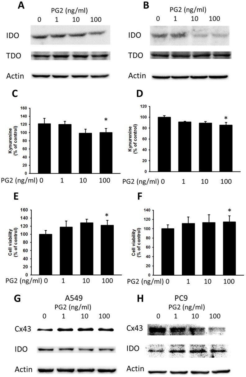
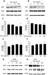
- Figure 4 The effects of PG2 on the expression and function of IDO. PG2 inhibited IDO protein expression in (A) B16F10 and (B) LL2 cells in a dose-dependent manner. After treatment with PG2 (0-100 ng/ml) for 24 h, the expression of IDO and TDO levels in B16F10 and LL2 cells were measured by Western blotting. The kynurenine assay was used for the kynurenine production in (C) B16F10 and (D) LL2 cells. The conditioned medium of (E) B16F10 and (F) LL2 after treated with indicated concentrations of PG2 for 24 h mixed with an equal amount of original medium. T cells were cultured in media conditioned from tumor cells for 72 h. The cell number were measured by staining with trypan blue. (n = 6, data are mean+- SD. * P < 0.05). PG2 induced Cx43 and inhibited IDO protein expression in (G) A549 and (H) PC9 cells in a dose-dependent manner. After treatment with PG2 (0-100 ng/ml) for 24 h, the expression of Cx43 and IDO levels in A549 and PC9 cells were measured by Western blotting.