 Explore
 Validate
 Learn
Learn Western blot
Antibody data
- Antibody Data
- Antigen structure
- References [2]
- Comments [0]
- Validations
- Western blot [1]
- Immunohistochemistry [4]
- Other assay [2]
Submit
Validation data
Reference
Comment
Report error
- Product number
- PA5-34376 - Provider product page

- Provider
- Invitrogen Antibodies
- Product name
- ZFX Polyclonal Antibody
- Antibody type
- Polyclonal
- Antigen
- Synthetic peptide
- Description
- A suggested positive control is human small intestine tissue lysate. PA5-34376 can be used with blocking peptide PEP-1419.
- Reactivity
- Human, Mouse, Rat
- Host
- Rabbit
- Isotype
- IgG
- Vial size
- 100 μg
- Concentration
- 1 mg/mL
- Storage
- Maintain refrigerated at 2-8°C for up to 3 months. For long term storage store at -20°C
Submitted references Transcriptomic profiling of neonatal mouse granulosa cells reveals new insights into primordial follicle activation†.
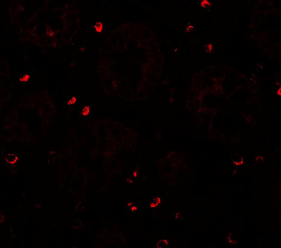
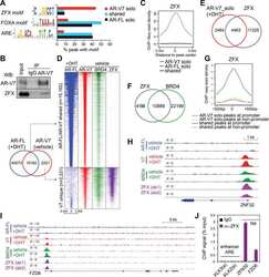
ZFX Mediates Non-canonical Oncogenic Functions of the Androgen Receptor Splice Variant 7 in Castrate-Resistant Prostate Cancer.
Ford EA, Frost ER, Beckett EL, Roman SD, McLaughlin EA, Sutherland JM
Biology of reproduction 2022 Mar 19;106(3):503-514
Biology of reproduction 2022 Mar 19;106(3):503-514
ZFX Mediates Non-canonical Oncogenic Functions of the Androgen Receptor Splice Variant 7 in Castrate-Resistant Prostate Cancer.
Cai L, Tsai YH, Wang P, Wang J, Li D, Fan H, Zhao Y, Bareja R, Lu R, Wilson EM, Sboner A, Whang YE, Zheng D, Parker JS, Earp HS, Wang GG
Molecular cell 2018 Oct 18;72(2):341-354.e6
Molecular cell 2018 Oct 18;72(2):341-354.e6
No comments: Submit comment
Supportive validation
- Submitted by
- Invitrogen Antibodies (provider)
- Main image
- Experimental details
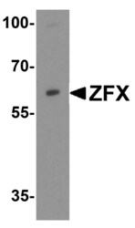
- Western Blot analysis of ZFX in Daudi cell lysate with ZFX Polyclonal Antibody (Product # PA5-34376) at 1 µg/mL.

Supportive validation
- Submitted by
- Invitrogen Antibodies (provider)
- Main image
- Experimental details
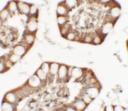
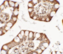
- Immunohistochemistry of ZFX in human small intestine tissue with ZFX Polyclonal Antibody (Product # PA5-34376) at 5 µg/mL.
- Submitted by
- Invitrogen Antibodies (provider)
- Main image
- Experimental details
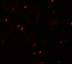
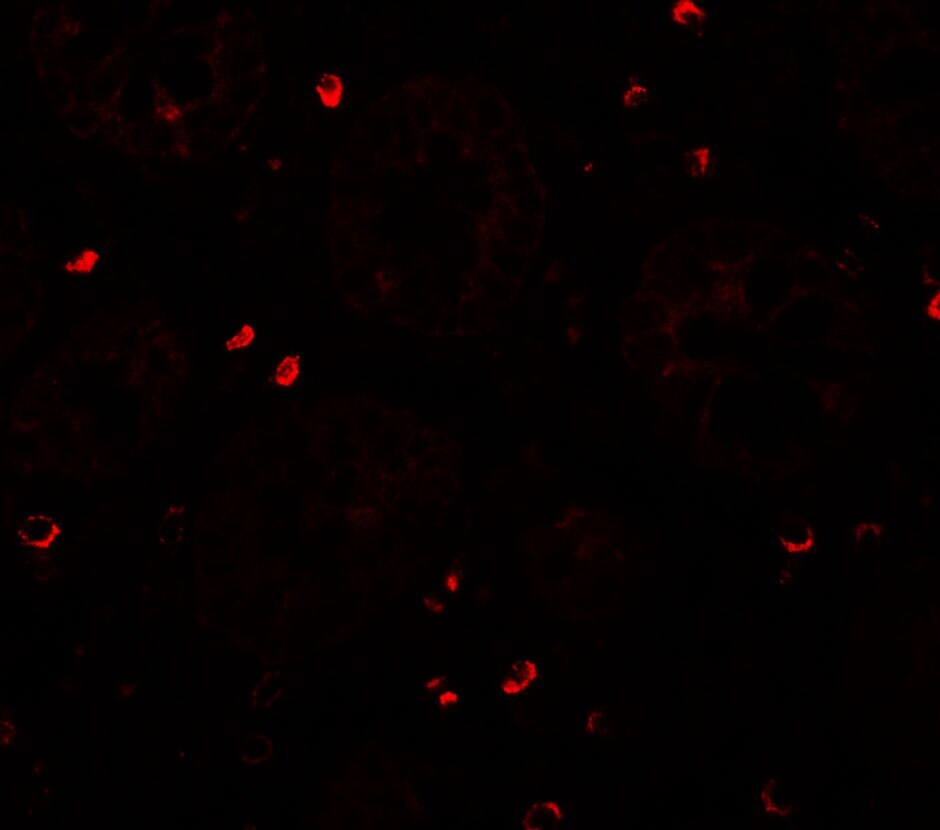
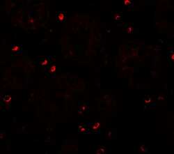
- Immunofluorescence of ZFX in human small intestine tissue with ZFX Polyclonal Antibody (Product # PA5-34376) at 20 µg/mL.
- Submitted by
- Invitrogen Antibodies (provider)
- Main image
- Experimental details
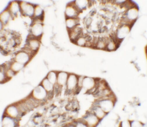
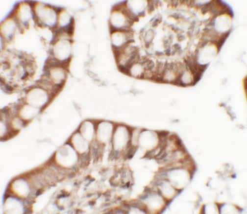
- Immunohistochemistry of ZFX in human small intestine tissue with ZFX Polyclonal Antibody (Product # PA5-34376) at 5 µg/mL.
- Submitted by
- Invitrogen Antibodies (provider)
- Main image
- Experimental details
- Immunofluorescence of ZFX in human small intestine tissue with ZFX Polyclonal Antibody (Product # PA5-34376) at 20 µg/mL.
Supportive validation
- Submitted by
- Invitrogen Antibodies (provider)
- Main image
- Experimental details
- NULL
- Submitted by
- Invitrogen Antibodies (provider)
- Main image
- Experimental details
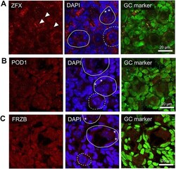
- Expression and localization of proteins of interest within the PND4 ovary. The in situ immunofluorescent expression of three proteins of interest (A-C) were explored in the neonatal mouse ovaries and were co-localized with a nuclear marker (DAPI, blue), a granulosa cell marker (GATA4 or FOXL2, green), and either (A) ZFX (B) POD1 or (C) FRZB in red. Representative images from PND4 selected as they include populations of primordial, activating and primary follicles. Representative images are indicative of n = 4-6 biological replicates of both PND1 and PND4 performed in triplicate. Images taken at 60x magnification, scale bars represent 20 mum with dotted circles outlining primordial follicles, solid lines outlining activating or primary follicles. Arrows indicate extracellular staining regions; asterisks indicate activating granulosa cells.
