Explore
 Validate
 Learn
Learn Western blot
 Immunocytochemistry
 Immunoprecipitation
Antibody data
- Antibody Data
- Antigen structure
- References [3]
- Comments [0]
- Validations
- Immunocytochemistry [4]
Submit
Validation data
Reference
Comment
Report error
- Product number
- PA1-841 - Provider product page

- Provider
- Invitrogen Antibodies
- Product name
- RIP140 Polyclonal Antibody
- Antibody type
- Polyclonal
- Antigen
- Synthetic peptide
- Description
- PA1-841 detects recombinant human Receptor-Interacting Protein 140 (RIP 140). PA1-841 has been successfully used in Western blot and immunoprecipitation procedures. By Western blot, this antibody detects two proteins at ~140 kDa representing human RIP 140 expressed in rabbit reticulocyte lysate. The PA1-841 immunogen is a synthetic peptide corresponding to residues G(718) N P T K G R V K K K E K T P L R D(735) of human RIP140. This immunizing peptide (Cat. # PEP-030) is available for use in neutralization and control experiments.
- Reactivity
- Human
- Host
- Rabbit
- Isotype
- IgG
- Vial size
- 100 μg
- Concentration
- 1 mg/mL
- Storage
- -20°C, Avoid Freeze/Thaw Cycles
Submitted references The role of receptor-interacting protein 140 in the accumulation of fat in ovariectomised rats.
Nuclear factor RIP140 modulates transcriptional activation by the estrogen receptor.
Nuclear factor RIP140 modulates transcriptional activation by the estrogen receptor.
Liu WH, Lee YM, Lam KK, Chen YF, Wang JJ, Yen MH, Cheng PY
Obesity surgery 2011 Jul;21(7):935-40
Obesity surgery 2011 Jul;21(7):935-40
Nuclear factor RIP140 modulates transcriptional activation by the estrogen receptor.
Cavaillès V, Dauvois S, L'Horset F, Lopez G, Hoare S, Kushner PJ, Parker MG
The EMBO journal 1995 Aug 1;14(15):3741-51
The EMBO journal 1995 Aug 1;14(15):3741-51
Nuclear factor RIP140 modulates transcriptional activation by the estrogen receptor.
Cavaillès V, Dauvois S, L'Horset F, Lopez G, Hoare S, Kushner PJ, Parker MG
The EMBO journal 1995 Aug 1;14(15):3741-51
The EMBO journal 1995 Aug 1;14(15):3741-51
No comments: Submit comment
Supportive validation
- Submitted by
- Invitrogen Antibodies (provider)
- Main image
- Experimental details
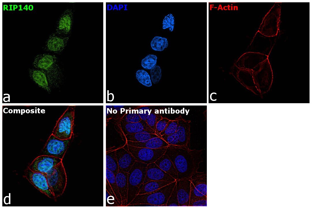
- Immunofluorescence analysis of RIP140 was performed using 70% confluent log phase MCF7 cells. The cells were fixed with 4% paraformaldehyde for 10 minutes, permeabilized with 0.1% Triton™ X-100 for 15 minutes, and blocked with 2% BSA for 45 minutes at room temperature. The cells were labeled with RIP140 Polyclonal Antibody (Product # PA1-841) at 1:200 dilution in 0.1% BSA, incubated at 4 degree celsius overnight and then labeled with Goat anti-Rabbit IgG (H+L) Highly Cross-Adsorbed Secondary Antibody, Alexa Fluor Plus 488 (Product # A32731), (1:3000 dilution), for 45 minutes at room temperature (Panel a: Green). Nuclei (Panel b:Blue) were stained with ProLong™ Diamond Antifade Mountant with DAPI (Product # P36962). F-actin (Panel c: Red) was stained with Rhodamine Phalloidin (Product # R415, 1:300). Panel d represents the merged image showing nucleus localization. Mild non specific cytoplasmic staining was also observed. Panel e represents control cells with no primary antibody to assess background. The images were captured at 60X magnification.

- Submitted by
- Invitrogen Antibodies (provider)
- Main image

- Experimental details
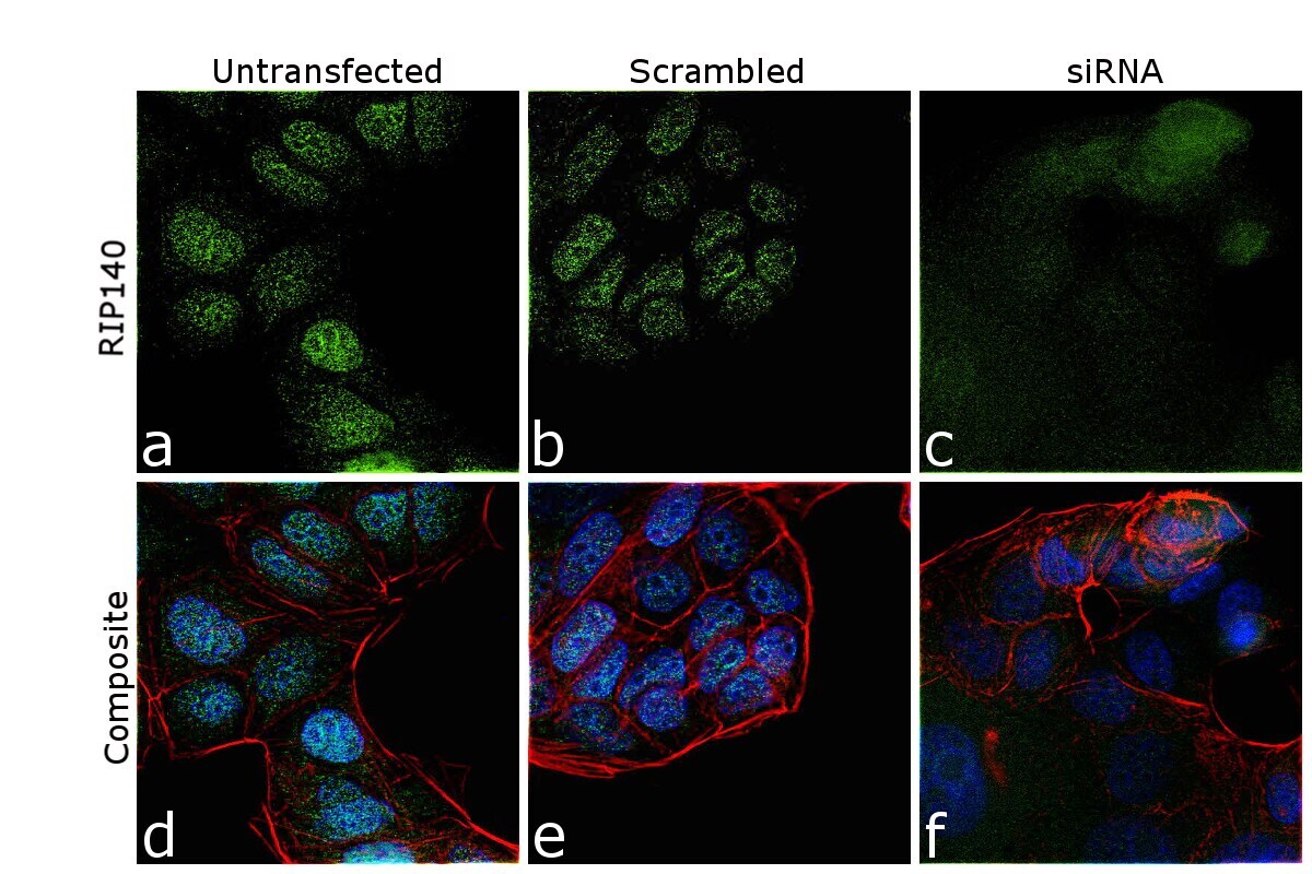
- Knockdown of RIP140 was achieved by transfecting MCF7 cells with RIP140 specific siRNA (Silencer® select Product # S15701, S15702). Immunofluorescence analysis was performed on untransfected MCF7 cells (panel a,d), transfected with non-specific scrambled siRNA (panels b,e) and transfected with RIP140 specific siRNA (panel c,f). Cells were fixed, permeabilized, and labelled with RIP140 Polyclonal Antibody (Product # PA1-841, 1:500 dilution) followed by Goat anti-Rabbit IgG (H+L) Highly Cross-Adsorbed Secondary Antibody, Alexa Fluor Plus 488 (Product # A32731), (1:3000dilution). Nuclei (blue) were stained using ProLong™ Diamond Antifade Mountant with DAPI (Product # P36962), and Rhodamine Phalloidin (Product # R415, 1:300) was used for cytoskeletal F-actin (Red) staining. Complete knockdown of RIP140 in siRNA transfected cells of specific signal was observed upon siRNA mediated knockdown (panel c,f) confirming specificity of the antibody to RIP140 (Green). The Images were captured at 60X magnification.

- Submitted by
- Invitrogen Antibodies (provider)
- Main image
- Experimental details
- Knockdown of RIP140 was achieved by transfecting MCF7 cells with RIP140 specific siRNA (Silencer® select Product # S15701, S15702). Immunofluorescence analysis was performed on untransfected MCF7 cells (panel a,d), transfected with non-specific scrambled siRNA (panels b,e) and transfected with RIP140 specific siRNA (panel c,f). Cells were fixed, permeabilized, and labelled with RIP140 Polyclonal Antibody (Product # PA1-841, 1:500 dilution) followed by Goat anti-Rabbit IgG (H+L) Highly Cross-Adsorbed Secondary Antibody, Alexa Fluor Plus 488 (Product # A32731), (1:3000dilution). Nuclei (blue) were stained using ProLong™ Diamond Antifade Mountant with DAPI (Product # P36962), and Rhodamine Phalloidin (Product # R415, 1:300) was used for cytoskeletal F-actin (Red) staining. Complete knockdown of RIP140 in siRNA transfected cells of specific signal was observed upon siRNA mediated knockdown (panel c,f) confirming specificity of the antibody to RIP140 (Green). The Images were captured at 60X magnification.
- Submitted by
- Invitrogen Antibodies (provider)
- Main image

- Experimental details
- Immunofluorescence analysis of RIP140 was performed using 70% confluent log phase MCF7 cells. The cells were fixed with 4% paraformaldehyde for 10 minutes, permeabilized with 0.1% Triton™ X-100 for 15 minutes, and blocked with 2% BSA for 45 minutes at room temperature. The cells were labeled with RIP140 Polyclonal Antibody (Product # PA1-841) at 1:200 dilution in 0.1% BSA, incubated at 4 degree celsius overnight and then labeled with Goat anti-Rabbit IgG (H+L) Highly Cross-Adsorbed Secondary Antibody, Alexa Fluor Plus 488 (Product # A32731), (1:3000 dilution), for 45 minutes at room temperature (Panel a: Green). Nuclei (Panel b:Blue) were stained with ProLong™ Diamond Antifade Mountant with DAPI (Product # P36962). F-actin (Panel c: Red) was stained with Rhodamine Phalloidin (Product # R415, 1:300). Panel d represents the merged image showing nucleus localization. Mild non specific cytoplasmic staining was also observed. Panel e represents control cells with no primary antibody to assess background. The images were captured at 60X magnification.