Explore
 Validate
 Learn
Learn Western blot
Antibody data
- Antibody Data
- Antigen structure
- References [0]
- Comments [0]
- Validations
- Western blot [2]
- Immunocytochemistry [2]
- Immunohistochemistry [8]
- Flow cytometry [1]
Submit
Validation data
Reference
Comment
Report error
- Product number
- R31053 - Provider product page

- Provider
- NSJ Bioreagents
- Product name
- CD31 Antibody
- Antibody type
- Polyclonal
- Antigen
- An amino acid sequence from the C-terminus of human CD31 (RKAVPDAVESRYSRTE) was used as the immunogen for this CD31 antibody.
- Description
- Antigen affinity purified antibody
- Reactivity
- Human
- Host
- Rabbit
- Conjugate
- Unconjugated
- Vial size
- 100 µg
- Concentration
- Lyophilized; resuspend with 200 ul for 0.5 mg/ml
- Storage
- After reconstitution, the CD31 antibody can be stored for up to one month at 4°C. For long-term, aliquot and store at -20°C. Avoid repeated freezing and thawing.
No comments: Submit comment
Supportive validation
- Submitted by
- NSJ Bioreagents (provider)
- Main image
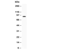
- Experimental details
- Western blot testing of CD31 antibody and human placenta lysate. Predicted size: 83-130KD depending on level of glycosylation

- Submitted by
- NSJ Bioreagents (provider)
- Main image
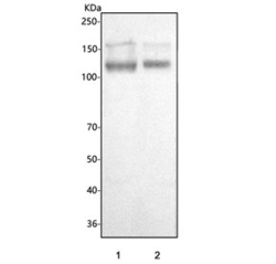
- Experimental details
- Western blot testing of human 1) HEL and 2) Jurkat cell lysate with CD31 antibody. Predicted molecular weight: 83-130 kDa depending on level of glycosylation.
Supportive validation
- Submitted by
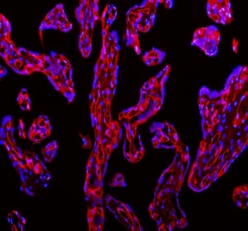
- NSJ Bioreagents (provider)
- Main image
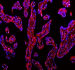
- Experimental details
- Immunofluorescent staining of FFPE human placental tissue with CD31 antibody (red) and DAPI nuclear stain (blue). HIER: steam section in pH8 EDTA buffer for 20 min.
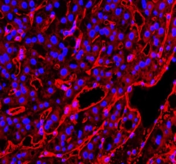
- Submitted by
- NSJ Bioreagents (provider)
- Main image
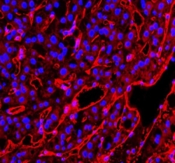
- Experimental details
- Immunofluorescent staining of FFPE human liver cancer tissue with CD31 antibody (red) and DAPI nuclear stain (blue). HIER: steam section in pH8 EDTA buffer for 20 min.
Supportive validation
- Submitted by
- NSJ Bioreagents (provider)
- Main image
- Experimental details
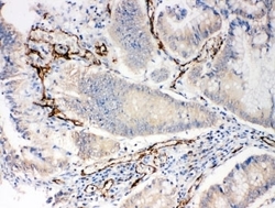
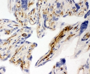
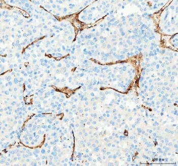
- IHC-P: CD31 antibody testing of human intestinal cancer tissue
- Submitted by
- NSJ Bioreagents (provider)
- Main image
- Experimental details
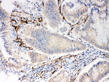
- IHC-P: CD31 antibody testing of human placenta tissue
- Submitted by
- NSJ Bioreagents (provider)
- Main image

- Experimental details
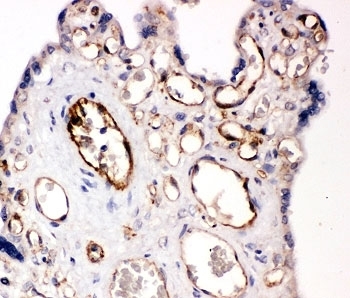
- IHC-F testing of CD31 antibody and human placenta tissue
- Submitted by
- NSJ Bioreagents (provider)
- Main image
- Experimental details
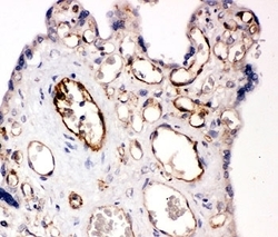
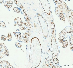
- IHC staining of FFPE human placental tissue with CD31 antibody. HIER: boil tissue sections in pH8 EDTA for 20 min and allow to cool before testing.
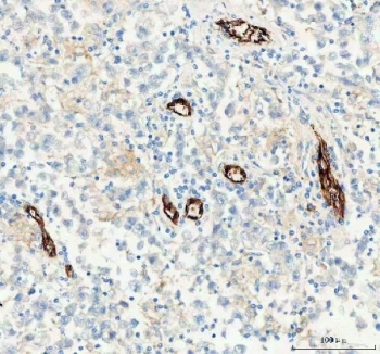
- Submitted by
- NSJ Bioreagents (provider)
- Main image

- Experimental details
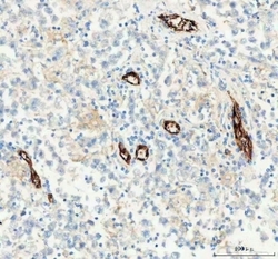
- IHC staining of FFPE human liver cancer tissue with CD31 antibody. HIER: boil tissue sections in pH8 EDTA for 20 min and allow to cool before testing.
- Submitted by
- NSJ Bioreagents (provider)
- Main image
- Experimental details
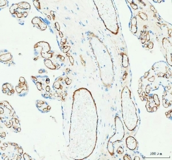
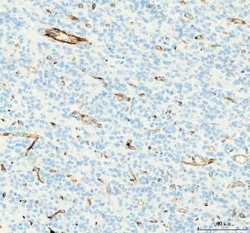
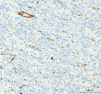
- IHC staining of FFPE human glioblastoma tissue with CD31 antibody. HIER: boil tissue sections in pH8 EDTA for 20 min and allow to cool before testing.
- Submitted by
- NSJ Bioreagents (provider)
- Main image
- Experimental details
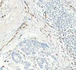
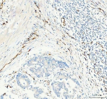
- IHC staining of FFPE human colorectal adenocarcinoma tissue with CD31 antibody. HIER: boil tissue sections in pH8 EDTA for 20 min and allow to cool before testing.
- Submitted by
- NSJ Bioreagents (provider)
- Main image
- Experimental details
- IHC staining of FFPE human testicular germ cell tumor tissue with CD31 antibody. HIER: boil tissue sections in pH8 EDTA for 20 min and allow to cool before testing.
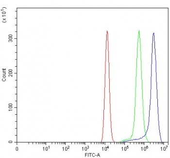
Supportive validation
- Submitted by
- NSJ Bioreagents (provider)
- Main image
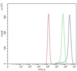
- Experimental details
- Flow cytometry testing of fixed and permeabilized human HEL cells with CD31 antibody at 1ug/million cells (blocked with goat sera); Red=cells alone, Green=isotype control, Blue= CD31 antibody.