Explore
 Validate
 Learn
Learn Western blot
 ELISA
ELISAAntibody data
- Antibody Data
- Antigen structure
- References [6]
- Comments [0]
- Validations
- Western blot [1]
Submit
Validation data
Reference
Comment
Report error
- Product number
- A01513-2 - Provider product page

- Provider
- Boster Biological Technology
- Product name
- Anti-CD31/Pecam1 Antibody Picoband™
- Antibody type
- Polyclonal
- Description
- Rabbit IgG polyclonal antibody for CD31/Pecam1 detection. Tested with WB, FCM, Direct ELISA in Mouse;Rat.
- Reactivity
- Mouse, Rat
- Host
- Rabbit
- Vial size
- 100μg/vial
- Concentration
- Add 0.2ml of distilled water will yield a concentration of 500ug/ml.
- Storage
- At -20°C for one year. After reconstitution, at 4°C for one month. It can also be aliquoted and stored frozen at -20°C for a longer time. Avoid repeated freezing and thawing.
- Handling
- Add 0.2ml of distilled water will yield a concentration of 500ug/ml.
Submitted references G-Quadruplex/Hemin DNAzyme-Functionalized Silver Nanoclusters with Synergistic Antibacterial and Wound Healing Capabilities for Infected Wound Management.
EPAC inhibitor suppresses angiogenesis and tumor growth of triple-negative breast cancer.
Exosomes Derived from E2F1(-/-) Adipose-Derived Stem Cells Promote Skin Wound Healing via miR-130b-5p/TGFBR3 Axis.
Antibacterial, conductive nanocomposite hydrogel based on dextran, carboxymethyl chitosan and chitosan oligosaccharide for diabetic wound therapy and health monitoring.
Angiogenesis-Browning Interplay Mediated by Asprosin-Knockout Contributes to Weight Loss in Mice with Obesity.
Performance of a multilayered small-diameter vascular scaffold dual-loaded with VEGF and PDGF.
Zhu J, Wen T, Qu S, Li Q, Liu B, Zhou W
Small (Weinheim an der Bergstrasse, Germany) 2024 Feb;20(8):e2307220
Small (Weinheim an der Bergstrasse, Germany) 2024 Feb;20(8):e2307220
EPAC inhibitor suppresses angiogenesis and tumor growth of triple-negative breast cancer.
Li Z, Liu Q, Cai Y, Ye N, He Z, Yao Y, Ding Y, Wang P, Qi C, Zheng L, Wang L, Zhou J, Zhang QQ
Biochimica et biophysica acta. Molecular basis of disease 2024 Apr;1870(4):167114
Biochimica et biophysica acta. Molecular basis of disease 2024 Apr;1870(4):167114
Exosomes Derived from E2F1(-/-) Adipose-Derived Stem Cells Promote Skin Wound Healing via miR-130b-5p/TGFBR3 Axis.
Yu H, Wu Y, Zhang B, Xiong M, Yi Y, Zhang Q, Wu M
International journal of nanomedicine 2023;18:6275-6292
International journal of nanomedicine 2023;18:6275-6292
Antibacterial, conductive nanocomposite hydrogel based on dextran, carboxymethyl chitosan and chitosan oligosaccharide for diabetic wound therapy and health monitoring.
Zhao N, Yuan W
International journal of biological macromolecules 2023 Dec 31;253(Pt 1):126625
International journal of biological macromolecules 2023 Dec 31;253(Pt 1):126625
Angiogenesis-Browning Interplay Mediated by Asprosin-Knockout Contributes to Weight Loss in Mice with Obesity.
Yin T, Chen S, Zeng G, Yuan W, Lu Y, Zhang Y, Huang Q, Xiong X, Xu B, Huang Q
International journal of molecular sciences 2022 Dec 18;23(24)
International journal of molecular sciences 2022 Dec 18;23(24)
Performance of a multilayered small-diameter vascular scaffold dual-loaded with VEGF and PDGF.
Han F, Jia X, Dai D, Yang X, Zhao J, Zhao Y, Fan Y, Yuan X
Biomaterials 2013 Oct;34(30):7302-13
Biomaterials 2013 Oct;34(30):7302-13
No comments: Submit comment
Supportive validation
- Submitted by
- Boster Biological Technology (provider)
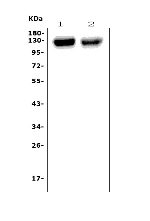
- Main image
- Experimental details
- Western blot analysis of Pecam1 using anti-Pecam1 antibody (A01513-2). Electrophoresis was performed on a 5-20% SDS-PAGE gel at 70V (Stacking gel) / 90V (Resolving gel) for 2-3 hours. The sample well of each lane was loaded with 50ug of sample under reducing conditions. Lane 1: rat lung tissue lysates, Lane 2: mouse lung tissue lysates. After Electrophoresis, proteins were transferred to a Nitrocellulose membrane at 150mA for 50-90 minutes. Blocked the membrane with 5% Non-fat Milk/ TBS for 1.5 hour at RT. The membrane was incubated with rabbit anti-Pecam1 antigen affinity purified polyclonal antibody (Catalog # A01513-2) at 0.25 μg/mL overnight at 4°C, then washed with TBS-0.1%Tween 3 times with 5 minutes each and probed with a goat anti-rabbit IgG-HRP secondary antibody at a dilution of 1:5000 for 1.5 hour at RT. The signal is developed using an Enhanced Chemiluminescent detection (ECL) kit (Catalog # EK1002) with Tanon 5200 system. A specific band was detected for Pecam1 at approximately 120-130KD. The expected band size for Pecam1 is at 82KD.
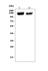
- Additional image