Explore
 Validate
 Learn
Learn Western blot
 Immunocytochemistry
Antibody data
- Antibody Data
- Antigen structure
- References [1]
- Comments [0]
- Validations
- Immunocytochemistry [1]
Submit
Validation data
Reference
Comment
Report error
- Product number
- HPA023057 - Provider product page

- Provider
- Atlas Antibodies
- Proper citation
- Atlas Antibodies Cat#HPA023057, RRID:AB_1852338
- Product name
- Anti-KIAA0753
- Antibody type
- Polyclonal
- Description
- Polyclonal Antibody against Human KIAA0753, Gene description: KIAA0753, Validated applications: ICC, IHC, WB, Uniprot ID: Q2KHM9, Storage: Store at +4°C for short term storage. Long time storage is recommended at -20°C.
- Reactivity
- Human
- Host
- Rabbit
- Conjugate
- Unconjugated
- Isotype
- IgG
- Vial size
- 100 µl
- Concentration
- 0.1 mg/ml
- Storage
- Store at +4°C for short term storage. Long time storage is recommended at -20°C.
- Handling
- The antibody solution should be gently mixed before use.
Submitted references Novel asymmetrically localizing components of human centrosomes identified by complementary proteomics methods
Jakobsen L, Vanselow K, Skogs M, Toyoda Y, Lundberg E, Poser I, Falkenby L, Bennetzen M, Westendorf J, Nigg E, Uhlen M, Hyman A, Andersen J
The EMBO Journal 2011;30(8):1520-1535
The EMBO Journal 2011;30(8):1520-1535
No comments: Submit comment
Supportive validation
- Submitted by
- Atlas Antibodies (provider)
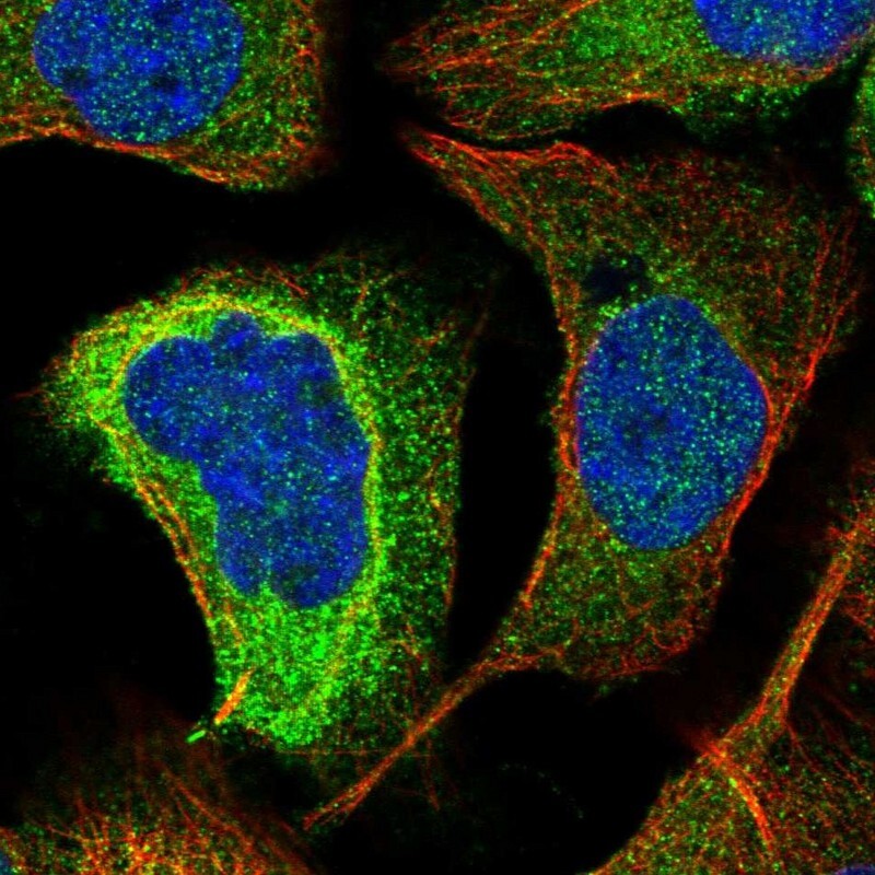
- Main image

- Experimental details
- Immunofluorescent staining of human cell line A-431 shows localization to cytosol & centrosome.
- Sample type
- Human