Explore
 Validate
 Learn
Learn10068-1-AP
antibody from Invitrogen Antibodies
Targeting: TUBB6
HsT1601, MGC4083
 Western blot Immunocytochemistry
 Immunoprecipitation Immunohistochemistry Flow cytometry Other assay
Antibody data
- Antibody Data
- Antigen structure
- References [0]
- Comments [0]
- Validations
- Western blot [8]
- Immunocytochemistry [3]
- Immunohistochemistry [6]
- Flow cytometry [1]
- Other assay [2]
Submit
Validation data
Reference
Comment
Report error
- Product number
- 10068-1-AP - Provider product page

- Provider
- Invitrogen Antibodies
- Product name
- beta Tubulin Polyclonal Antibody
- Antibody type
- Polyclonal
- Antigen
- Other
- Reactivity
- Human, Mouse, Rat
- Host
- Rabbit
- Isotype
- IgG
- Vial size
- 150 µL
- Concentration
- 0.13 mg/mL
- Storage
- -20°C
No comments: Submit comment
Supportive validation
- Submitted by
- Invitrogen Antibodies (provider)
- Main image
- Experimental details
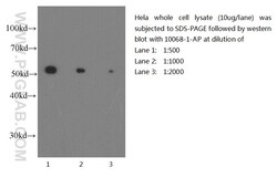
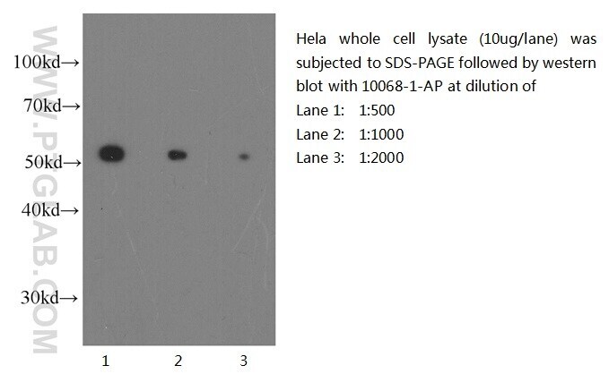
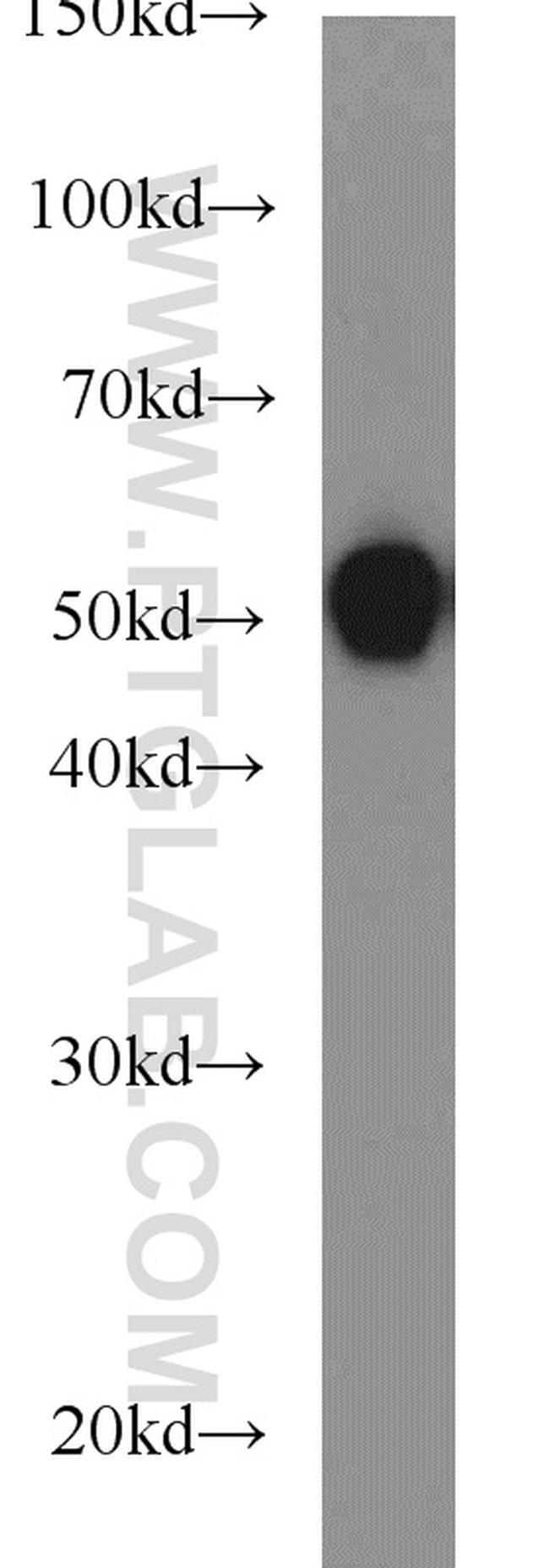
- Western blot of Hela cell with anti-Tubulin-Beta (10068-1-AP) at various dilutions.
- Submitted by
- Invitrogen Antibodies (provider)
- Main image
- Experimental details
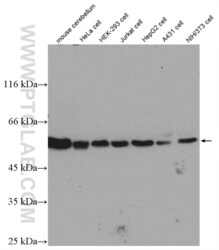
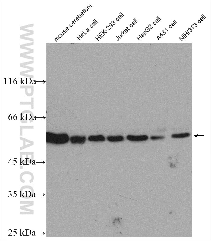
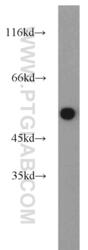
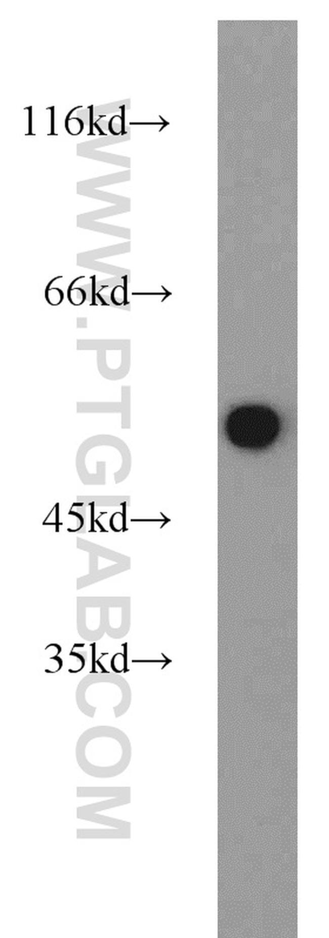
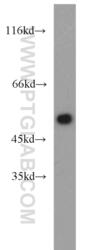
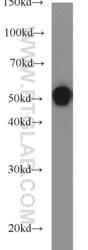
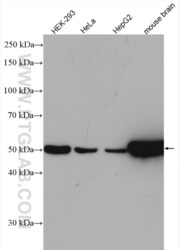
- Various lysates were subjected to SDS PAGE followed by western blot with 10068-1-AP (beta Tubulin antibody) at dilution of 1:1000 incubated at room temperature for 1.5 hours.
- Submitted by
- Invitrogen Antibodies (provider)
- Main image
- Experimental details
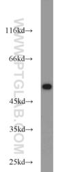
- HepG2 cells were subjected to SDS PAGE followed by western blot with 10068-1-AP (beta Tubulin antibody) at dilution of 1:1000 incubated at room temperature for 1.5 hours.
- Submitted by
- Invitrogen Antibodies (provider)
- Main image

- Experimental details
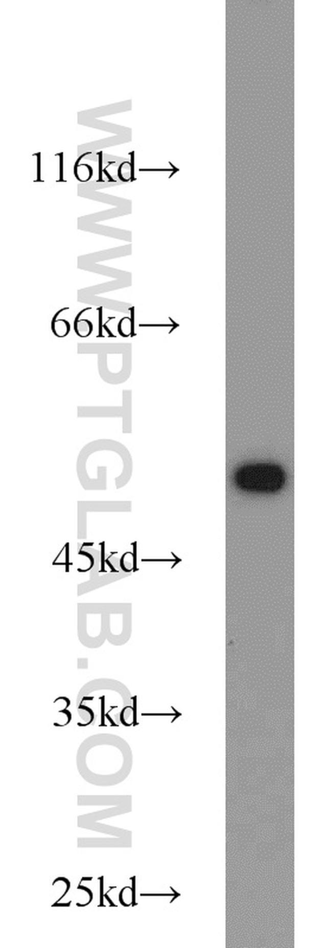
- COLO 320 cells were subjected to SDS PAGE followed by western blot with 10068-1-AP (beta Tubulin antibody) at dilution of 1:1000 incubated at room temperature for 1.5 hours.
- Submitted by
- Invitrogen Antibodies (provider)
- Main image
- Experimental details
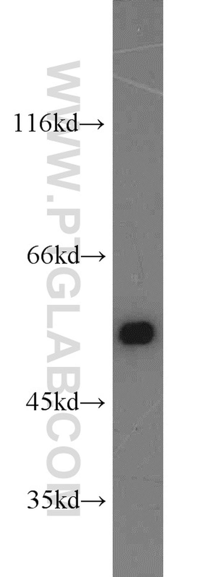
- Jurkat cells were subjected to SDS PAGE followed by western blot with 10068-1-AP (beta Tubulin antibody) at dilution of 1:1000 incubated at room temperature for 1.5 hours.
- Submitted by
- Invitrogen Antibodies (provider)
- Main image
- Experimental details
- HeLa cells were subjected to SDS PAGE followed by western blot with 10068-1-AP (beta Tubulin antibody) at dilution of 1:1000 incubated at room temperature for 1.5 hours.

- Submitted by
- Invitrogen Antibodies (provider)
- Main image
- Experimental details
- Neuro-2a cells were subjected to SDS PAGE followed by western blot with 10068-1-AP (beta Tubulin antibody) at dilution of 1:1000 incubated at room temperature for 1.5 hours.
- Submitted by
- Invitrogen Antibodies (provider)
- Main image
- Experimental details
- Various lysates were subjected to SDS PAGE followed by western blot with 10068-1-AP (beta Tubulin antibody) at dilution of 1:4000 incubated at room temperature for 1.5 hours.

Supportive validation
- Submitted by
- Invitrogen Antibodies (provider)
- Main image
- Experimental details
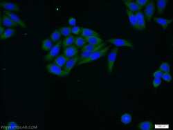
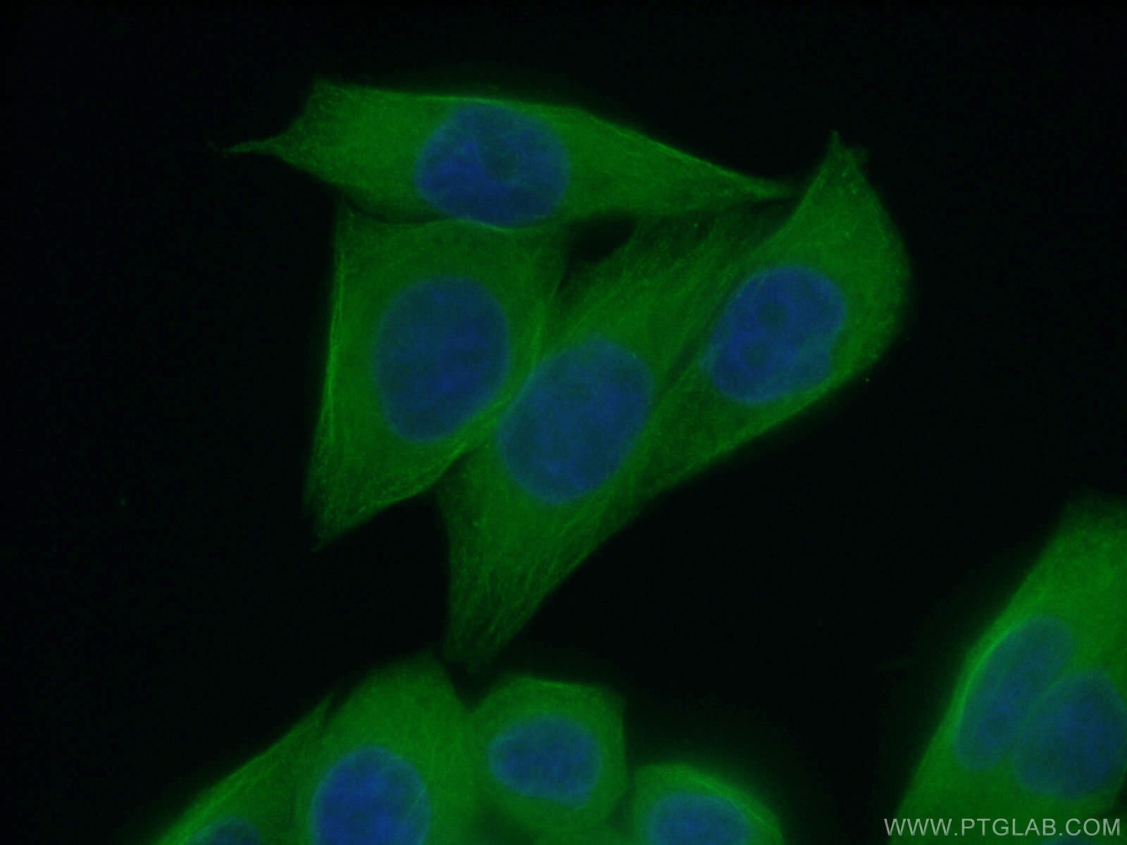
- Immunofluorescent analysis of HeLa cells using 10068-1-AP (beta Tubulin antibody) at dilution of 1:25 and Alexa Fluor 488-conjugated AffiniPure Goat Anti-Rabbit IGG (H+L).

- Submitted by
- Invitrogen Antibodies (provider)
- Main image
- Experimental details
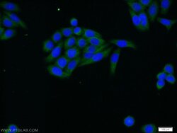
- Immunofluorescent analysis of HeLa cells using 10068-1-AP (beta Tubulin antibody) at dilution of 1:25 and Alexa Fluor 488-conjugated AffiniPure Goat Anti-Rabbit IGG (H+L).

- Submitted by
- Invitrogen Antibodies (provider)
- Main image

- Experimental details
- Immunofluorescent analysis of (4% PFA) fixed HeLa cells using 10068-1-AP (beta Tubulin antibody) at dilution of 1:50 and Alexa Fluor 488-Conjugated AffiniPure Goat Anti-Rabbit IgG (H+L).
Supportive validation
- Submitted by
- Invitrogen Antibodies (provider)
- Main image
- Experimental details
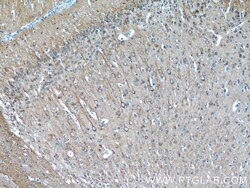
- Immunohistochemistry of paraffin-embedded mouse brain tissue slide using 10068-1-AP ( beta Tubulin antibody at dilution of 1:50 (under 10x lens).
- Submitted by
- Invitrogen Antibodies (provider)
- Main image
- Experimental details
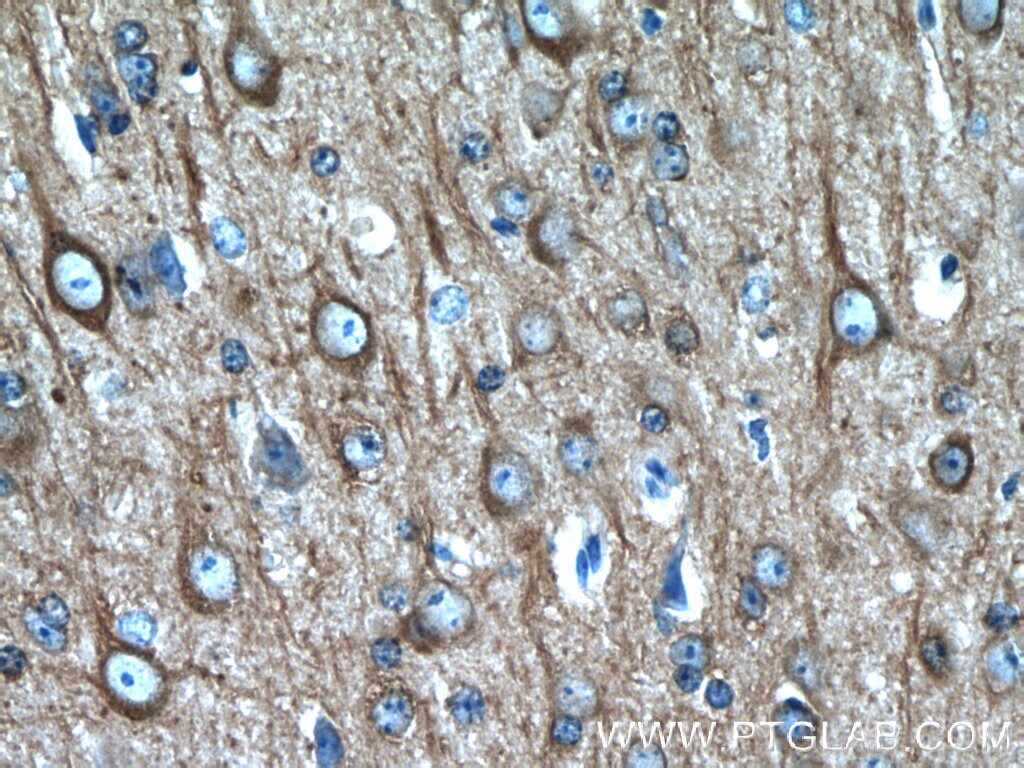
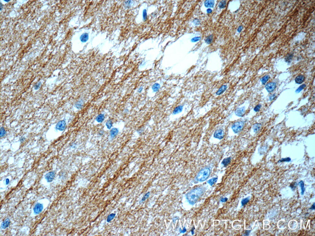
- Immunohistochemistry of paraffin-embedded mouse brain tissue slide using 10068-1-AP ( beta Tubulin antibody at dilution of 1:50 (under 40x lens).
- Submitted by
- Invitrogen Antibodies (provider)
- Main image
- Experimental details
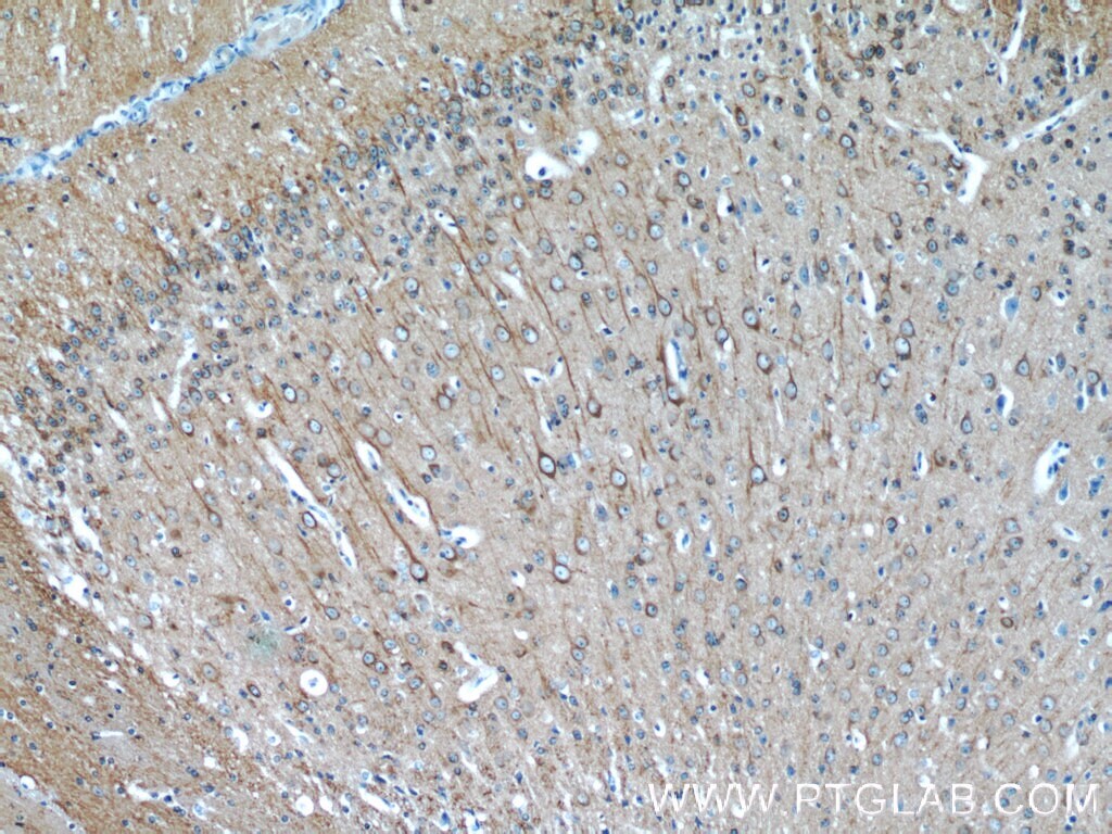
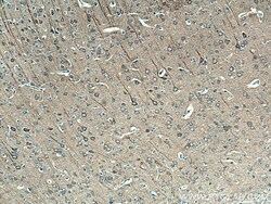
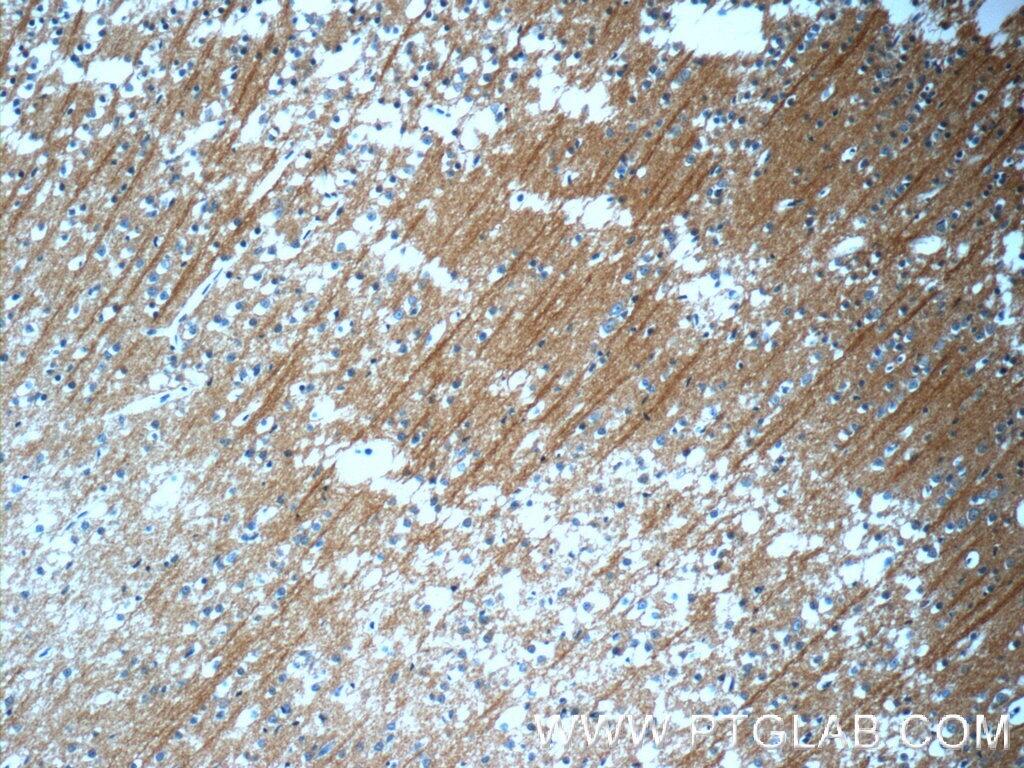
- Immunohistochemistry of paraffin-embedded rat brain tissue slide using 10068-1-AP (beta Tubulin antibody) at dilution of 1:200 (under 10x lens).
- Submitted by
- Invitrogen Antibodies (provider)
- Main image

- Experimental details
- Immunohistochemistry of paraffin-embedded rat brain tissue slide using 10068-1-AP (beta Tubulin antibody) at dilution of 1:200 (under 40x lens).
- Submitted by
- Invitrogen Antibodies (provider)
- Main image
- Experimental details
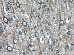
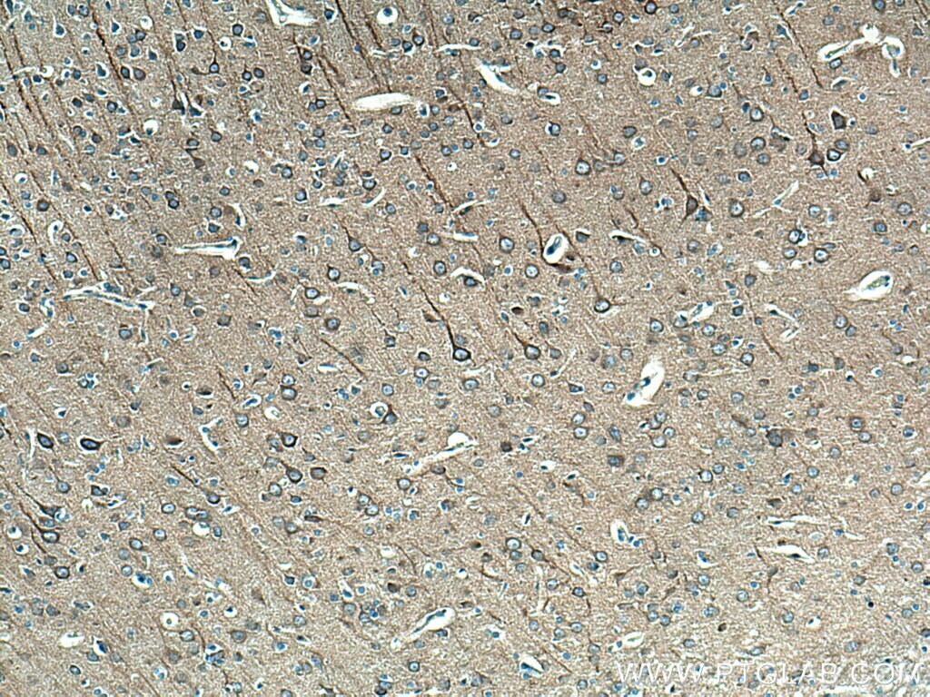
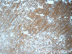
- Immunohistochemistry of paraffin-embedded human brain tissue slide using 10068-1-AP (beta Tubulin antibody at dilution of 1:50 (under 10x lens).
- Submitted by
- Invitrogen Antibodies (provider)
- Main image

- Experimental details
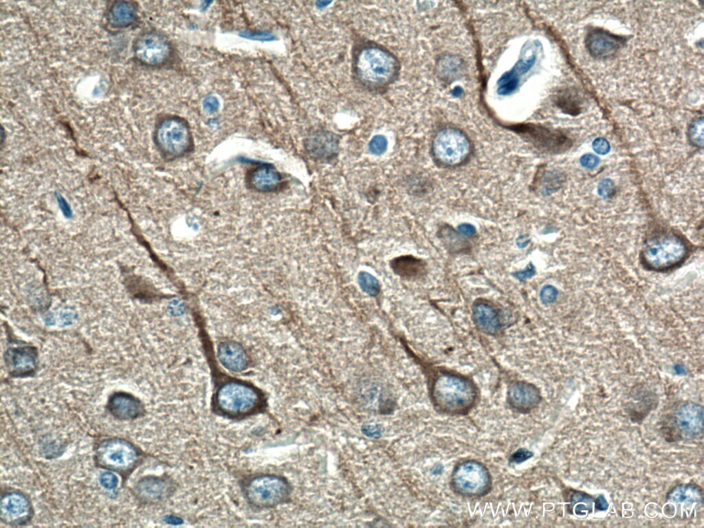
- Immunohistochemistry of paraffin-embedded human brain tissue slide using 10068-1-AP (beta Tubulin antibody at dilution of 1:50 (under 40x lens).
Supportive validation
- Submitted by
- Invitrogen Antibodies (provider)
- Main image

- Experimental details
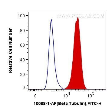
- 1X10^6 HeLa cells were intracellularly stained with 0.4 µg Anti-Human Beta Tubulin (Product # 10068-1-AP) and CoraLite®488-Conjugated AffiniPure Goat Anti-Rabbit IgG(H+L) at dilution 1:1,000 (red), or 0.4 µg Control Antibody. Cells were fixed with 4% PFA and permeabilized with Flow Cytometry Perm Buffer (PF00011-C).
Supportive validation
- Submitted by
- Invitrogen Antibodies (provider)
- Main image
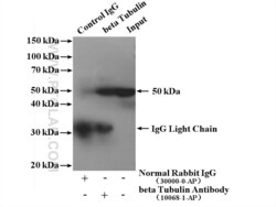
- Experimental details
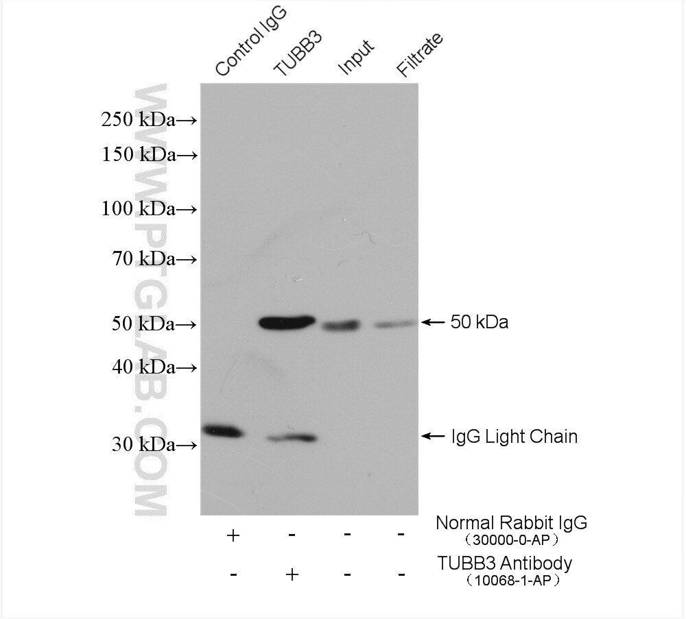
- IP result of anti-beta Tubulin (IP:10068-1-AP, 4ug; Detection:10068-1-AP 1:1000) with mouse brain tissue lysate 3160 ug.

- Submitted by
- Invitrogen Antibodies (provider)
- Main image

- Experimental details
- IP result of anti-beta Tubulin (IP:10068-1-AP, 4 ug; Detection:10068-1-AP 1:5000) with HEK-293 cells lysate 2320 ug.