 Explore
 Validate
 Learn
Learn Western blot
Antibody data
- Antibody Data
- Antigen structure
- References [17]
- Comments [0]
- Validations
- Western blot [4]
- Immunocytochemistry [1]
- Immunohistochemistry [3]
- Flow cytometry [1]
- Other assay [4]
Submit
Validation data
Reference
Comment
Report error
- Product number
- PA5-16863 - Provider product page

- Provider
- Invitrogen Antibodies
- Product name
- beta Tubulin Polyclonal Antibody
- Antibody type
- Polyclonal
- Antigen
- Synthetic peptide
- Description
- PA5-16863 targets Tubulin Beta in IHC (P), WB, and IP applications and shows reactivity with Amphibian, Bovine, Chicken, Fungi, Gerbil, Guinea Pig, Human, mouse, Porcine, Rat, Sea Urchin, and Yeast samples.
- Concentration
- 0.09 mg/mL
Submitted references CTP synthase is essential for early endosperm development by regulating nuclei spacing.
The Gastroprotective Effect of Naringenin against Ethanol-Induced Gastric Ulcers in Mice through Inhibiting Oxidative and Inflammatory Responses.
Cigarette Smoke Activates NOTCH3 to Promote Goblet Cell Differentiation in Human Airway Epithelial Cells.
A mouse model of Huntington's disease shows altered ultrastructure of transverse tubules in skeletal muscle fibers.
Prognostic value of SH3PXD2B (Tks4) in human hepatocellular carcinoma: a combined multi-omics and experimental study.
Study of the cytological features of bone marrow mesenchymal stem cells from patients with neuromyelitis optica.
Aged blood factors decrease cellular responses associated with delayed gingival wound repair.
Ribosome-bound Pub1 modulates stop codon decoding during translation termination in yeast.
Genetic Evidence for the Role of the Vacuole in Supplying Secretory Organelles with Ca2+ in Hansenula polymorpha.
Fetal inhibition of inflammation improves disease phenotypes in harlequin ichthyosis.
Phospholipase C-related catalytically inactive protein (PRIP) controls KIF5B-mediated insulin secretion.
N-Cadherin is a prospective cell surface marker of human mesenchymal stem cells that have high ability for cardiomyocyte differentiation.
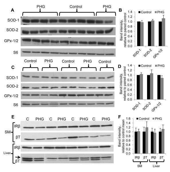
Chronic pulsatile hyperglycemia reduces insulin secretion and increases accumulation of reactive oxygen species in fetal sheep islets.
Expression of proto-oncogene KIT is up-regulated in subset of human meningiomas.
Heterogeneous time-dependent response of adipose tissue during the development of cancer cachexia.
A subset of human gliomas shows over-expression of KIT without its amplification.
Neurite outgrowth of mature retinal ganglion cells and PC12 cells requires activity of CK1δ and CK1ε.
Yoon J, Cho LH, Kim SR, Tun W, Peng X, Pasriga R, Moon S, Hong WJ, Ji H, Jung KH, Jeon JS, An G
Plant biotechnology journal 2021 Nov;19(11):2177-2191
Plant biotechnology journal 2021 Nov;19(11):2177-2191
The Gastroprotective Effect of Naringenin against Ethanol-Induced Gastric Ulcers in Mice through Inhibiting Oxidative and Inflammatory Responses.
Li WS, Lin SC, Chu CH, Chang YK, Zhang X, Lin CC, Tung YT
International journal of molecular sciences 2021 Nov 5;22(21)
International journal of molecular sciences 2021 Nov 5;22(21)
Cigarette Smoke Activates NOTCH3 to Promote Goblet Cell Differentiation in Human Airway Epithelial Cells.
Bodas M, Moore AR, Subramaniyan B, Georgescu C, Wren JD, Freeman WM, Brown BR, Metcalf JP, Walters MS
American journal of respiratory cell and molecular biology 2021 Apr;64(4):426-440
American journal of respiratory cell and molecular biology 2021 Apr;64(4):426-440
A mouse model of Huntington's disease shows altered ultrastructure of transverse tubules in skeletal muscle fibers.
Romer SH, Metzger S, Peraza K, Wright MC, Jobe DS, Song LS, Rich MM, Foy BD, Talmadge RJ, Voss AA
The Journal of general physiology 2021 Apr 5;153(4)
The Journal of general physiology 2021 Apr 5;153(4)
Prognostic value of SH3PXD2B (Tks4) in human hepatocellular carcinoma: a combined multi-omics and experimental study.
Kui X, Wang Y, Zhang C, Li H, Li Q, Ke Y, Wang L
BMC medical genomics 2021 Apr 28;14(1):115
BMC medical genomics 2021 Apr 28;14(1):115
Study of the cytological features of bone marrow mesenchymal stem cells from patients with neuromyelitis optica.
Yang C, Yang Y, Ma L, Zhang GX, Shi FD, Yan Y, Chang G
International journal of molecular medicine 2019 Mar;43(3):1395-1405
International journal of molecular medicine 2019 Mar;43(3):1395-1405
Aged blood factors decrease cellular responses associated with delayed gingival wound repair.
Saldías MP, Fernández C, Morgan A, Díaz C, Morales D, Jaña F, Gómez A, Silva A, Briceño F, Oyarzún A, Maldonado F, Cerda O, Smith PC, Cáceres M
PloS one 2017;12(9):e0184189
PloS one 2017;12(9):e0184189
Ribosome-bound Pub1 modulates stop codon decoding during translation termination in yeast.
Urakov VN, Mitkevich OV, Safenkova IV, Ter-Avanesyan MD
The FEBS journal 2017 Jun;284(12):1914-1930
The FEBS journal 2017 Jun;284(12):1914-1930
Genetic Evidence for the Role of the Vacuole in Supplying Secretory Organelles with Ca2+ in Hansenula polymorpha.
Fokina AV, Chechenova MB, Karginov AV, Ter-Avanesyan MD, Agaphonov MO
PloS one 2015;10(12):e0145915
PloS one 2015;10(12):e0145915
Fetal inhibition of inflammation improves disease phenotypes in harlequin ichthyosis.
Cottle DL, Ursino GM, Ip SC, Jones LK, Ditommaso T, Hacking DF, Mangan NE, Mellett NA, Henley KJ, Sviridov D, Nold-Petry CA, Nold MF, Meikle PJ, Kile BT, Smyth IM
Human molecular genetics 2015 Jan 15;24(2):436-49
Human molecular genetics 2015 Jan 15;24(2):436-49
Phospholipase C-related catalytically inactive protein (PRIP) controls KIF5B-mediated insulin secretion.
Asano S, Nemoto T, Kitayama T, Harada K, Zhang J, Harada K, Tanida I, Hirata M, Kanematsu T
Biology open 2014 May 8;3(6):463-74
Biology open 2014 May 8;3(6):463-74
N-Cadherin is a prospective cell surface marker of human mesenchymal stem cells that have high ability for cardiomyocyte differentiation.
Ishimine H, Yamakawa N, Sasao M, Tadokoro M, Kami D, Komazaki S, Tokuhara M, Takada H, Ito Y, Kuno S, Yoshimura K, Umezawa A, Ohgushi H, Asashima M, Kurisaki A
Biochemical and biophysical research communications 2013 Sep 6;438(4):753-9
Biochemical and biophysical research communications 2013 Sep 6;438(4):753-9
Chronic pulsatile hyperglycemia reduces insulin secretion and increases accumulation of reactive oxygen species in fetal sheep islets.
Green AS, Chen X, Macko AR, Anderson MJ, Kelly AC, Hart NJ, Lynch RM, Limesand SW
The Journal of endocrinology 2012 Mar;212(3):327-42
The Journal of endocrinology 2012 Mar;212(3):327-42
Expression of proto-oncogene KIT is up-regulated in subset of human meningiomas.
Saini M, Jha AN, Abrari A, Ali S
BMC cancer 2012 Jun 6;12:212
BMC cancer 2012 Jun 6;12:212
Heterogeneous time-dependent response of adipose tissue during the development of cancer cachexia.
Batista ML Jr, Neves RX, Peres SB, Yamashita AS, Shida CS, Farmer SR, Seelaender M
The Journal of endocrinology 2012 Dec;215(3):363-73
The Journal of endocrinology 2012 Dec;215(3):363-73
A subset of human gliomas shows over-expression of KIT without its amplification.
Saini M, Jha AN, Abrari A, Ali S
Gene 2012 Apr 15;497(2):155-63
Gene 2012 Apr 15;497(2):155-63
Neurite outgrowth of mature retinal ganglion cells and PC12 cells requires activity of CK1δ and CK1ε.
Bischof J, Müller A, Fänder M, Knippschild U, Fischer D
PloS one 2011;6(6):e20857
PloS one 2011;6(6):e20857
No comments: Submit comment
Supportive validation
- Submitted by
- Invitrogen Antibodies (provider)
- Main image

- Experimental details
- Western blot of Tubulin Beta using Tubulin Beta Polyclonal Antibody (Product # PA5-16863) on MCF-7 Cells.

- Submitted by
- Invitrogen Antibodies (provider)
- Main image

- Experimental details
- Western blot analysis was performed on whole cell extracts (30µg lysate) of A549 (Lane 1), COS-7 (Lane 2), MDCK (Lane 3), C2C12 (Lane 4), MDA-MB-231 (Lane 5) PC-12 (Lane 6), RSC96 (Lane 7), tissue extracts of Mouse Lung (Lane 8), Rat Stomach (Lane 9) and Rat Brain (Lane 10). The blot was probed with Anti-beta Tubulin Rabbit Polyclonal Antibody (Product # PA5-16863, 1:1000 dilution) and detected by chemiluminescence using Goat anti-Rabbit IgG (H+L) Superclonal™ Secondary Antibody, HRP conjugate (Product # A28177, 0.25µg/mL, 1:4000 dilution). A 52 kDa band corresponding to beta Tubulin was observed across the cell lines and tissues tested. Known quantity of protein samples were electrophoresed using Novex® NuPAGE® 4-12 % Bis-Tris gel (Product # NP0321BOX), XCell SureLock™ Electrophoresis System (Product # EI0002) and Novex® Sharp Pre-Stained Protein Standard (Product # LC5800). Resolved proteins were then transferred onto a nitrocellulose membrane with iBlot® 2 Dry Blotting System (Product # IB21001). The membrane was probed with the relevant primary and secondary Antibody following blocking with 5 % skimmed milk. Chemiluminescent detection was performed using Pierce™ ECL Western Blotting Substrate (Product # 32106).

- Submitted by
- Invitrogen Antibodies (provider)
- Main image

- Experimental details
- Western blot analysis was performed on whole cell extracts (30 µg lysate) of MDA-MB-231 (Lane 1), MCF-7 (Lane 2), A431 (Lane 3), A549 (Lane 4), HEK 293 (Lane 5) and HeLa (Lane6). The blots were probed with Anti-Tubulin Beta Rabbit Polyclonal Antibody (Product # PA5-16863, 1:1000 dilution) and detected by chemiluminescence Goat Anti-Rabbit IgG (H+L) Secondary Antibody, HRP conjµgate (Product # G-21234, 1:5000 dilution). A 50 kDa band corresponding to Tubulin was observed across cell lines tested. Known quantity of protein samples were electrophoresed using Novex® NuPAGE® 12 % Bis-Tris gel (Product # NP0341BOX), XCell SureLock™ Electrophoresis System (Product # EI0002) and Novex® Sharp Pre-Stained Protein Standard (Product # LC5800). Resolved proteins were then transferred onto a nitrocellulose membrane with iBlot® 2 Dry Blotting System (Product # IB21001). The membrane was probed with the relevant primary and secondary Antibody following blocking with 5 % skimmed milk. Chemiluminescent detection was performed using Pierce™ ECL Western Blotting Substrate (Product # 32106).

- Submitted by
- Invitrogen Antibodies (provider)
- Main image

- Experimental details
- Western blot analysis was performed on whole cell extracts (30µg lysate) of A549 (Lane 1), COS-7 (Lane 2), MDCK (Lane 3), C2C12 (Lane 4), MDA-MB-231 (Lane 5) PC-12 (Lane 6), RSC96 (Lane 7), tissue extracts of Mouse Lung (Lane 8), Rat Stomach (Lane 9) and Rat Brain (Lane 10). The blot was probed with Anti-beta Tubulin Rabbit Polyclonal Antibody (Product # PA5-16863, 1:1000 dilution) and detected by chemiluminescence using Goat anti-Rabbit IgG (H+L) Superclonal™ Secondary Antibody, HRP conjugate (Product # A28177, 0.25µg/mL, 1:4000 dilution). A 52 kDa band corresponding to beta Tubulin was observed across the cell lines and tissues tested. Known quantity of protein samples were electrophoresed using Novex® NuPAGE® 4-12 % Bis-Tris gel (Product # NP0321BOX), XCell SureLock™ Electrophoresis System (Product # EI0002) and Novex® Sharp Pre-Stained Protein Standard (Product # LC5800). Resolved proteins were then transferred onto a nitrocellulose membrane with iBlot® 2 Dry Blotting System (Product # IB21001). The membrane was probed with the relevant primary and secondary Antibody following blocking with 5 % skimmed milk. Chemiluminescent detection was performed using Pierce™ ECL Western Blotting Substrate (Product # 32106).

Supportive validation
- Submitted by
- Invitrogen Antibodies (provider)
- Main image

- Experimental details
- Immunofluorescence analysis of Tubulin Beta was performed using 70% confluent log phase NIH/3T3 cells. The cells were fixed with 4% paraformaldehyde for 10 minutes, permeabilized with 0.1% Triton™ X-100 for 10 minutes, and blocked with 2% BSA for 1 hour at room temperature. The cells were labeled with Tubulin Beta Rabbit Polyclonal Antibody (Product # PA5-16863) at 2 µg/mL in 0.1% BSA and incubated for 3 hours at room temperature and then labeled with Goat anti-Rabbit IgG (H+L) Superclonal™ Secondary Antibody, Alexa Fluor® 488 conjugate (Product # A27034) a dilution of 1:2000 for 45 minutes at room temperature (Panel a: green). Nuclei (Panel b: blue) were stained with SlowFade® Gold Antifade Mountant with DAPI (Product # S36938). F-actin (Panel c: red) was stained with Alexa Fluor® 555 Rhodamine Phalloidin (Product # R415, 1:300). Panel d represents the merged image showing cytoplasmic localization. Panel e shows the no primary antibody control. The images were captured at 60X magnification.

Supportive validation
- Submitted by
- Invitrogen Antibodies (provider)
- Main image

- Experimental details
- Formalin-fixed, paraffin-embedded human lung stained with Tubulin beta antibody using peroxidase-conjugate and AEC. Note cytoplasmic staining of ciliated epithelium of bronchioles.

- Submitted by
- Invitrogen Antibodies (provider)
- Main image

- Experimental details
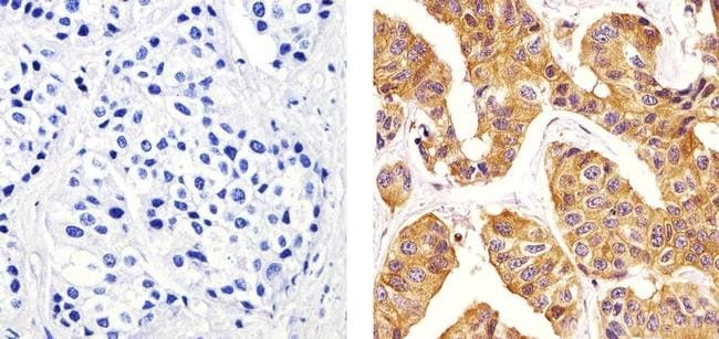
- Immunohistochemistry analysis of Tubulin Beta showing staining in the cytoplasm of paraffin-embedded human colon carcinoma (right) compared to a negative control without primary antibody (left). To expose target proteins, antigen retrieval was performed using 10mM sodium citrate (pH 6.0), microwaved for 8-15 min. Following antigen retrieval, tissues were blocked in 3% H2O2-methanol for 15 min at room temperature, washed with ddH2O and PBS, and then probed with a Tubulin Beta Rabbit Polyclonal Antibody (Product # PA5-16863) diluted in 3% BSA-PBS at a dilution of 1:20 for 1 hour at 37°C in a humidified chamber. Tissues were washed extensively in PBST and detection was performed using an HRP-conjugated secondary antibody followed by colorimetric detection using a DAB kit. Tissues were counterstained with hematoxylin and dehydrated with ethanol and xylene to prep for mounting.

- Submitted by
- Invitrogen Antibodies (provider)
- Main image

- Experimental details
- Immunohistochemistry analysis of Tubulin Beta showing staining in the cytoplasm of paraffin-embedded human breast carcinoma (right) compared to a negative control without primary antibody (left). To expose target proteins, antigen retrieval was performed using 10mM sodium citrate (pH 6.0), microwaved for 8-15 min. Following antigen retrieval, tissues were blocked in 3% H2O2-methanol for 15 min at room temperature, washed with ddH2O and PBS, and then probed with a Tubulin Beta Rabbit Polyclonal Antibody (Product # PA5-16863) diluted in 3% BSA-PBS at a dilution of 1:20 for 1 hour at 37°C in a humidified chamber. Tissues were washed extensively in PBST and detection was performed using an HRP-conjugated secondary antibody followed by colorimetric detection using a DAB kit. Tissues were counterstained with hematoxylin and dehydrated with ethanol and xylene to prep for mounting.
Supportive validation
- Submitted by
- Invitrogen Antibodies (provider)
- Main image
- Experimental details
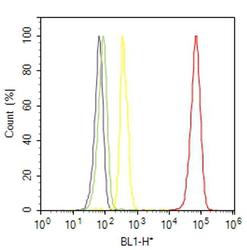
- Flow cytometry analysis of Tubulin Beta was done on HeLa cells. Cells were fixed with 70% ethanol for 10 minutes, permeabilized with 0.25% Triton™ X-100 for 20 minutes, and blocked with 5% BSA for 30 minutes at room temperature. Cells were labeled with Tubulin Beta Rabbit Polyclonal Antibody (PA516863, red histogram) or with rabbit isotype control (yellow histogram) at 3-5 ug/million cells in 2.5% BSA. After incubation at room temperature for 2 hours, the cells were labeled with Alexa Fluor® 488 Goat Anti-Rabbit Secondary Antibody (A11008) at a dilution of 1:400 for 30 minutes at room temperature. The representative 10,000 cells were acquired and analyzed for each sample using an Attune® Acoustic Focusing Cytometer. The purple histogram represents unstained control cells and the green histogram represents no-primary-antibody control.

Supportive validation
- Submitted by
- Invitrogen Antibodies (provider)
- Main image

- Experimental details
- NULL
- Submitted by
- Invitrogen Antibodies (provider)
- Main image

- Experimental details
- Fig 6 Effect of inactivation of PMC1 on the level of Pmr1 in the ret1-27 mutant strain and in the strain bearing the RET1 wild-type allele. Proteins from cell lysates were resolved by SDS PAGE and transferred to nitrocellulose membrane, which was then divided in two parts at the level of the 80 kDa marker band. The upper part was stained with antiserum against H . polymorpha Pmr1, while the lower part was stained with antibody against tubulin used as a loading control. ret1-27 pmc1-Delta , the 64MA70QA-Deltapmc strain; pmc1-Delta , the 64MA70Q-RET-Deltapmc strain; ret1-27 , the 64MA70QAL strain; WT and WT 1/2, undiluted and two-fold diluted sample of the 64MA70QL-RET strain, respectively.

- Submitted by
- Invitrogen Antibodies (provider)
- Main image

- Experimental details
- Fig. 3 SH3PXD2B silencing inhibits the invasion, but not proliferation of Hep3B and Huh7 cells. Hep3B and Huh7 cells were transduced with lentivirus for expression of SH3PXD2B-specific shRNA or control shRNA (Scr). The relative levels of SH3PXD2B to the control beta-tubulin protein expression in different groups of Hep3B ( a , b ) and Huh7 ( c , d ) cells were determined by Western blot. e The dynamic proliferation of Hep3B and Huh7 cells was determined at the indicated time points. f , g The invasion of different groups of Hep3B and Huh7 cells was examined by transwell invasion assays. h - j The formation of invadopodia in different groups of Hep3B and Huh7 cells was examined by immunofluorescence assays. k - m The function of invadopodia in different groups of Hep3B and Huh7 cells was examined by in situ zymography. A total of 150 cells per group were analyzed by two researchers in a blinded manner. Data are representative images or expressed as the mean +- SD of each group from three separate experiments. Bar scale in F = 100 mum. Bar scales in H and K = 20 mum

- Submitted by
- Invitrogen Antibodies (provider)
- Main image

- Experimental details
- Figure 8. Bin1 protein levels in muscles of R6/2 and control mice . (A-D) Western blots for Bin1 protein in the quadriceps femoris (A and B) and the gastrocnemius (C and D) of age-matched late-stage (10-13 wk of age) control (WT) and R6/2 mice. (A and C) Representative Western blots for Bin1 as well as beta-tubulin and GAPDH for normalization. Equal amounts of protein were loaded per lane (50 ug). (B and D) Bin1 band intensities normalized to beta-tubulin and GAPDH levels (relative to control) for the quadriceps femoris (B) and gastrocnemius (D). For A and B (quadriceps femoris), n = 4/group. For C and D (gastrocnemius), n = 6/group. *, Significantly different from control at P < 0.05. AU, arbitrary units.
