 Explore
 Validate
 Learn
Learn Western blot
 Immunocytochemistry
Antibody data
- Antibody Data
- Antigen structure
- References [1]
- Comments [0]
- Validations
- Immunocytochemistry [1]
Submit
Validation data
Reference
Comment
Report error
- Product number
- MAB2547 - Provider product page

- Provider
- R&D Systems
- Product name
- Human FGF-20 Antibody
- Antibody type
- Monoclonal
- Description
- Protein A or G purified from hybridoma culture supernatant. Detects human FGF-20 in direct ELISAs and Western blots. In direct ELISAs and Western blots, no cross-reactivity with recombinant human (rh) FGF-3, -4, -5, -6, -7, -9, -10, -11, -12, -16, -17, -18, -19, -23, recombinant mouse FGF-8b, -8c, -15, -23, rhFGF acidic, or rhFGF basic is observed.
- Reactivity
- Human
- Host
- Rat
- Conjugate
- Unconjugated
- Antigen sequence
Q9NP95- Isotype
- IgG
- Antibody clone number
- 272317
- Vial size
- 100 ug
- Concentration
- LYOPH
- Storage
- Use a manual defrost freezer and avoid repeated freeze-thaw cycles. 12 months from date of receipt, -20 to -70 °C as supplied. 1 month, 2 to 8 °C under sterile conditions after reconstitution. 6 months, -20 to -70 °C under sterile conditions after reconstitution.
Submitted references Variation in the miRNA-433 binding site of FGF20 confers risk for Parkinson disease by overexpression of alpha-synuclein.
Wang G, van der Walt JM, Mayhew G, Li YJ, Züchner S, Scott WK, Martin ER, Vance JM
American journal of human genetics 2008 Feb;82(2):283-9
American journal of human genetics 2008 Feb;82(2):283-9
No comments: Submit comment
Supportive validation
- Submitted by
- R&D Systems (provider)
- Main image
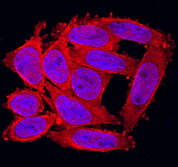
- Experimental details
- FGF-20 in SW480 Human Cell Line. FGF-20 was detected in immersion fixed SW480 human colorectal adenocarcinoma cell line using Rat Anti-Human FGF-20 Monoclonal Antibody (Catalog # MAB2547) at 10 µg/mL for 3 hours at room temperature. Cells were stained using the NorthernLights™ 557-conjugated Anti-Rat IgG Secondary Antibody (red; Catalog # NL013) and counterstained with DAPI (blue). Specific staining was localized to cytoplasm. View our protocol for Fluorescent ICC Staining of Cells on Coverslips.
