 Explore
 Validate
 Learn
Learn Western blot
 Immunoprecipitation
 Immunohistochemistry
Antibody data
- Antibody Data
- Antigen structure
- References [1]
- Comments [0]
- Validations
- Immunohistochemistry [1]
Submit
Validation data
Reference
Comment
Report error
- Product number
- MA3-003 - Provider product page

- Provider
- Invitrogen Antibodies
- Product name
- BAP31 Monoclonal Antibody (CC-4)
- Antibody type
- Monoclonal
- Antigen
- Other
- Description
- MA3-003 detects BAP31 from human, non-human primate, bovine and hamster samples. MA3-003 has been successfully used in Western blot, immunofluorescent, and immunohistochemical procedures. By Western blot, this antibody detects a 28-kDa protein corresponding to human BAP31. The MA3-003 antigen is solubilized protein from human neuroendocrine cell line IMR-32, followed by an extract of the small lung human carcinoma cell line SCC-9. MA3-003 binds to a more proximal region of BAP31 (amino acids 123-229). This sequence is conserved in non-human primate, bovine and hamster species.
- Reactivity
- Human, Bovine, Hamster
- Host
- Rat
- Isotype
- IgG
- Antibody clone number
- CC-4
- Vial size
- 100 μL
- Concentration
- Conc. Not Determined
- Storage
- -20°C, Avoid Freeze/Thaw Cycles
Submitted references Association of BAP31 with CD11b/CD18. Potential role in intracellular trafficking of CD11b/CD18 in neutrophils.
Zen K, Utech M, Liu Y, Soto I, Nusrat A, Parkos CA
The Journal of biological chemistry 2004 Oct 22;279(43):44924-30
The Journal of biological chemistry 2004 Oct 22;279(43):44924-30
No comments: Submit comment
Supportive validation
- Submitted by
- Invitrogen Antibodies (provider)
- Main image
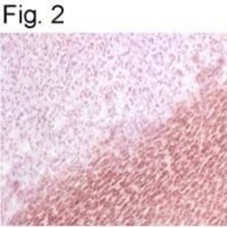
- Experimental details
- Immunohistochemical staining of BAP31 in baboon pituitary gland using Product # MA3-003.
