 Explore
 Validate
 Learn
Learn Western blot
 Immunoprecipitation
Antibody data
- Antibody Data
- Antigen structure
- References [0]
- Comments [0]
- Validations
- Western blot [3]
- Immunocytochemistry [1]
Submit
Validation data
Reference
Comment
Report error
- Product number
- PA5-22045 - Provider product page

- Provider
- Invitrogen Antibodies
- Product name
- Anti-TUBB8 Polyclonal Antibody
- Antibody type
- Polyclonal
- Antigen
- Synthetic peptide
- Description
- Recommended positive controls: A431, H1299, Neuro 2A, GL261, C8D30, NIH-3T3, BCL-1, Raw264.7, C2C12. Store product as a concentrated solution. Centrifuge briefly prior to opening the vial.
- Reactivity
- Human, Mouse, Rat
- Host
- Rabbit
- Isotype
- IgG
- Vial size
- 100 µL
- Concentration
- 1 mg/mL
- Storage
- Store at 4°C short term. For long term storage, store at -20°C, avoiding freeze/thaw cycles.
No comments: Submit comment
Supportive validation
- Submitted by
- Invitrogen Antibodies (provider)
- Main image
- Experimental details
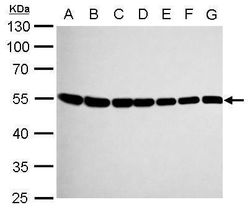
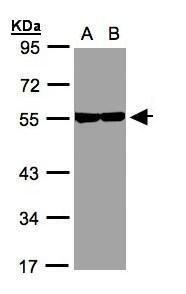
- Western blot analysis of Tubulin beta-8 using A) 30 µg Neuro2A whole cell lysate (B) 30 µg GL261 whole cell lysate (C) 30 µg C8D30 whole cell lysate (D) 30 µg NIH-3T3 whole cell lysate (E) 30 µg BCL-1 whole cell lysate (F) 30 µg Raw264.7 whole cell lysate and G) 30 µg C2C12 whole cell lysate. Samples were loaded onto a 10% SDS-PAGE gel and probed with a Tubulin beta-8 polyclonal antibody (Product # PA5-22045) at a dilution of 1:1000.
- Submitted by
- Invitrogen Antibodies (provider)
- Main image
- Experimental details
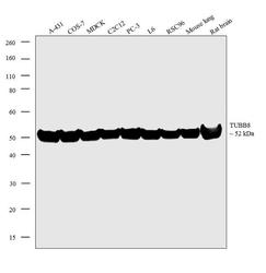
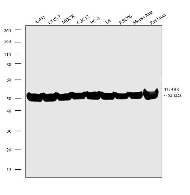
- Western blot analysis was performed on whole cell extracts (30µg lysate) of A-431 (Lane 1), COS-7 (Lane 2), MDCK (Lane 3), C2C12 (Lane 4), PC-3 (Lane 5), L6 (Lane 6), RSC96 (Lane 7), and tissue extracts (30µg lysate) of mouse lung (Lane 8), and rat brain (Lane 9). The blots were probed with Anti-TUBB8 Rabbit Polyclonal Antibody (Product # PA5-22045, 1:1000 dilution) and detected by chemiluminescence using Goat anti-Rabbit IgG (H+L) Superclonal™ Secondary Antibody, HRP conjugate (Product # A27036, 0.25µg/mL, 1:4000 dilution). A 52 kDa band corresponding to TUBB8 was observed across the cell lines and tissues tested. Known quantity of protein samples were electrophoresed using Novex® NuPAGE® 4-12 % Bis-Tris gel (Product # NP0321BOX), XCell SureLock™ Electrophoresis System (Product # EI0002) and Novex® Sharp Pre-Stained Protein Standard (Product # LC5800). Resolved proteins were then transferred onto a nitrocellulose membrane with iBlot® 2 Dry Blotting System (Product # IB21001). The membrane was probed with the relevant primary and secondary Antibody following blocking with 5 % skimmed milk. Chemiluminescent detection was performed using Pierce™ ECL Western Blotting Substrate (Product # 32106).
- Submitted by
- Invitrogen Antibodies (provider)
- Main image
- Experimental details
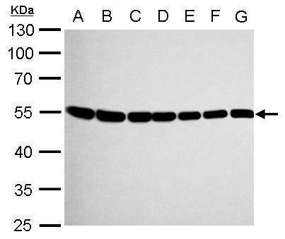
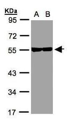
- Western Blot analysis of TUBB8 was performed by separating 30 µg of various whole cell extracts by 10% SDS PAGE. Proteins were transferred to a membrane and probed with a TUBB8 Polyclonal Antibody (Product # PA5-22045) at a dilution of 1:1000. A:A431, B:H1299.
Supportive validation
- Submitted by
- Invitrogen Antibodies (provider)
- Main image
- Experimental details
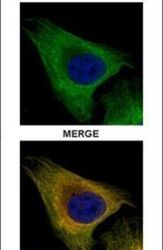
- Immunofluorescent analysis of Tubulin beta-8 in paraformaldehyde-fixed U2OS cells using a Tubulin beta-8 polyclonal antibody (Product # PA5-22045) (Green) at a 1:500 dilution. Alpha-tubulin filaments were labeled with Product # PA5-29281 (Red) at a 1:2000.
