 Explore
 Validate
 Learn
Learn Western blot
 ELISA
ELISAAntibody data
- Antibody Data
- Antigen structure
- References [1]
- Comments [0]
- Validations
- Western blot [1]
- Immunohistochemistry [3]
Submit
Validation data
Reference
Comment
Report error
- Product number
- LS-C19035 - Provider product page

- Provider
- LSBio
- Proper citation
- LifeSpan Cat#LS-C19035, RRID:AB_2092975
- Product name
- DLL4 Antibody (Internal) LS-C19035
- Antibody type
- Polyclonal
- Description
- Affinity purified
- Reactivity
- Human
- Host
- Rabbit
- Isotype
- IgG
- Storage
- Short term: store at 4°C. Long term: store at -20°C. Avoid freeze-thaw cycles.
Submitted references Cholera toxin disrupts barrier function by inhibiting exocyst-mediated trafficking of host proteins to intestinal cell junctions.
Guichard A, Cruz-Moreno B, Aguilar B, van Sorge NM, Kuang J, Kurkciyan AA, Wang Z, Hang S, Pineton de Chambrun GP, McCole DF, Watnick P, Nizet V, Bier E
Cell host & microbe 2013 Sep 11;14(3):294-305
Cell host & microbe 2013 Sep 11;14(3):294-305
No comments: Submit comment
Enhanced validation
- Submitted by
- LSBio (provider)
- Enhanced method
- Genetic validation
- Main image
- Experimental details
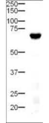
- Western Blot - Anti-Delta-4 Antibody. Western blot of Affinity Purified anti-Delta-4 antibody shows detection of a 74-kD band corresponding to Delta-4 in a lysate prepared from mouse pancreatic tissue. Approximately 20 ug of lysate was run on SDS-PAGE and transferred onto nitrocellulose followed by reaction with a 1:500 dilution of anti-Delta-4 antibody. Detection occurred using a 1:5000 dilution of HRP-labeled Goat anti-Rabbit IgG for 1 hour at room temperature. A chemiluminescence system was used for signal detection (Roche) using a 3 min exposure time.

Supportive validation
- Submitted by
- LSBio (provider)
- Enhanced method
- Genetic validation
- Main image
- Experimental details
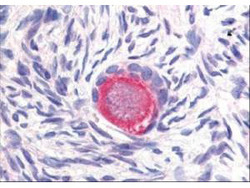
- Anti-Delta-4 Antibody - Immunohistochemistry. Affinity Purified anti-Delta-4 antibody was used at 20 ug/ml to detect Delta-4 in a variety of tissues including colon, liver, skeletal muscle, ovary, pancreas, prostate, testes, thymus, tonsil and uterus. In contrast to reported findings, no staining was observed in vascular tissue. This image shows Delta-4 staining of human ovary. Tissue was formalin-fixed and paraffin embedded. Personal Communication, Tina Roush, LifeSpanBiosciences, Seattle, WA.
- Submitted by
- LSBio (provider)
- Enhanced method
- Genetic validation
- Main image

- Experimental details
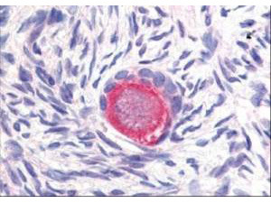
- Anti-Delta-4 Antibody - Immunohistochemistry. Affinity Purified anti-Delta-4 antibody was used at 20 ug/ml to detect Delta-4 in a variety of tissues including colon, liver, skeletal muscle, ovary, pancreas, prostate, testes, thymus, tonsil and uterus. In contrast to reported findings, no staining was observed in vascular tissue. This image shows Delta-4 staining of human ovary. Tissue was formalin-fixed and paraffin embedded. Personal Communication, Tina Roush, LifeSpanBiosciences, Seattle, WA.

- Submitted by
- LSBio (provider)
- Enhanced method
- Genetic validation
- Main image

- Experimental details
- Anti-Delta-4 Antibody - Immunohistochemistry. Affinity Purified anti-Delta-4 antibody was used at 20 ug/ml to detect Delta-4 in a variety of tissues including colon, liver, skeletal muscle, ovary, pancreas, prostate, testes, thymus, tonsil and uterus. In contrast to reported findings, no staining was observed in vascular tissue. This image shows Delta-4 staining of human ovary. Tissue was formalin-fixed and paraffin embedded. Personal Communication, Tina Roush, LifeSpanBiosciences, Seattle, WA.
