Explore
 Validate
 Learn
Learn Western blot
 Immunohistochemistry
Antibody data
- Antibody Data
- Antigen structure
- References [19]
- Comments [0]
- Validations
- Western blot [1]
- Immunocytochemistry [1]
Submit
Validation data
Reference
Comment
Report error
- Product number
- AF1389 - Provider product page

- Provider
- R&D Systems
- Product name
- Mouse DLL4 Antibody
- Antibody type
- Polyclonal
- Description
- Antigen Affinity-purified. Detects mouse DLL4 in direct ELISAs and Western blots. In direct ELISAs, approximately 50% cross-reactivity with recombinant human DLL4 is observed.
- Reactivity
- Mouse
- Host
- Goat
- Conjugate
- Unconjugated
- Antigen sequence
BAB18580- Isotype
- IgG
- Vial size
- 100 ug
- Concentration
- LYOPH
- Storage
- Use a manual defrost freezer and avoid repeated freeze-thaw cycles. 12 months from date of receipt, -20 to -70 °C as supplied. 1 month, 2 to 8 °C under sterile conditions after reconstitution. 6 months, -20 to -70 °C under sterile conditions after reconstitution.
Submitted references iSuRe-Cre is a genetic tool to reliably induce and report Cre-dependent genetic modifications.
Venous identity requires BMP signalling through ALK3.
MPDZ promotes DLL4-induced Notch signaling during angiogenesis.
Lymphatic deletion of calcitonin receptor-like receptor exacerbates intestinal inflammation.
The endothelial transcription factor ERG mediates Angiopoietin-1-dependent control of Notch signalling and vascular stability.
Data showing proliferation and differentiation of intestinal epithelial cells under targeted depletion of Notch ligands in mouse intestine.
Blood flow controls bone vascular function and osteogenesis.
Notch Signaling Regulates the Homeostasis of Tissue-Restricted Innate-like T Cells.
Sox7, Sox17, and Sox18 Cooperatively Regulate Vascular Development in the Mouse Retina.
Slit2 signaling through Robo1 and Robo2 is required for retinal neovascularization.
Selective neuronal lineages derived from Dll4-expressing progenitors/precursors in the retina and spinal cord.
Interleukin-6 Stimulates Defective Angiogenesis.
Endothelial Notch activity promotes angiogenesis and osteogenesis in bone.
Neuroligin 1 induces blood vessel maturation by cooperating with the α6 integrin.
Fbxw7 controls angiogenesis by regulating endothelial Notch activity.
Notch-dependent VEGFR3 upregulation allows angiogenesis without VEGF-VEGFR2 signalling.
Delta1 expression, cell cycle exit, and commitment to a specific secretory fate coincide within a few hours in the mouse intestinal stem cell system.
Therapeutic efficacy of a DNA vaccine targeting the endothelial tip cell antigen delta-like ligand 4 in mammary carcinoma.
Blocking VEGFR-3 suppresses angiogenic sprouting and vascular network formation.
Fernández-Chacón M, Casquero-García V, Luo W, Francesca Lunella F, Ferreira Rocha S, Del Olmo-Cabrera S, Benedito R
Nature communications 2019 May 22;10(1):2262
Nature communications 2019 May 22;10(1):2262
Venous identity requires BMP signalling through ALK3.
Neal A, Nornes S, Payne S, Wallace MD, Fritzsche M, Louphrasitthiphol P, Wilkinson RN, Chouliaras KM, Liu K, Plant K, Sholapurkar R, Ratnayaka I, Herzog W, Bond G, Chico T, Bou-Gharios G, De Val S
Nature communications 2019 Jan 28;10(1):453
Nature communications 2019 Jan 28;10(1):453
MPDZ promotes DLL4-induced Notch signaling during angiogenesis.
Tetzlaff F, Adam MG, Feldner A, Moll I, Menuchin A, Rodriguez-Vita J, Sprinzak D, Fischer A
eLife 2018 Apr 5;7
eLife 2018 Apr 5;7
Lymphatic deletion of calcitonin receptor-like receptor exacerbates intestinal inflammation.
Davis RB, Kechele DO, Blakeney ES, Pawlak JB, Caron KM
JCI insight 2017 Mar 23;2(6):e92465
JCI insight 2017 Mar 23;2(6):e92465
The endothelial transcription factor ERG mediates Angiopoietin-1-dependent control of Notch signalling and vascular stability.
Shah AV, Birdsey GM, Peghaire C, Pitulescu ME, Dufton NP, Yang Y, Weinberg I, Osuna Almagro L, Payne L, Mason JC, Gerhardt H, Adams RH, Randi AM
Nature communications 2017 Jul 11;8:16002
Nature communications 2017 Jul 11;8:16002
Data showing proliferation and differentiation of intestinal epithelial cells under targeted depletion of Notch ligands in mouse intestine.
Nakata T, Shimizu H, Nagata S, Ito G, Fujii S, Suzuki K, Kawamoto A, Ishibashi F, Kuno R, Anzai S, Murano T, Mizutani T, Oshima S, Tsuchiya K, Nakamura T, Hozumi K, Watanabe M, Okamoto R
Data in brief 2017 Feb;10:551-556
Data in brief 2017 Feb;10:551-556
Blood flow controls bone vascular function and osteogenesis.
Ramasamy SK, Kusumbe AP, Schiller M, Zeuschner D, Bixel MG, Milia C, Gamrekelashvili J, Limbourg A, Medvinsky A, Santoro MM, Limbourg FP, Adams RH
Nature communications 2016 Dec 6;7:13601
Nature communications 2016 Dec 6;7:13601
Notch Signaling Regulates the Homeostasis of Tissue-Restricted Innate-like T Cells.
Chennupati V, Koch U, Coutaz M, Scarpellino L, Tacchini-Cottier F, Luther SA, Radtke F, Zehn D, MacDonald HR
Journal of immunology (Baltimore, Md. : 1950) 2016 Aug 1;197(3):771-82
Journal of immunology (Baltimore, Md. : 1950) 2016 Aug 1;197(3):771-82
Sox7, Sox17, and Sox18 Cooperatively Regulate Vascular Development in the Mouse Retina.
Zhou Y, Williams J, Smallwood PM, Nathans J
PloS one 2015;10(12):e0143650
PloS one 2015;10(12):e0143650
Slit2 signaling through Robo1 and Robo2 is required for retinal neovascularization.
Rama N, Dubrac A, Mathivet T, Ní Chárthaigh RA, Genet G, Cristofaro B, Pibouin-Fragner L, Ma L, Eichmann A, Chédotal A
Nature medicine 2015 May;21(5):483-91
Nature medicine 2015 May;21(5):483-91
Selective neuronal lineages derived from Dll4-expressing progenitors/precursors in the retina and spinal cord.
Zou M, Luo H, Xiang M
Developmental dynamics : an official publication of the American Association of Anatomists 2015 Jan;244(1):86-97
Developmental dynamics : an official publication of the American Association of Anatomists 2015 Jan;244(1):86-97
Interleukin-6 Stimulates Defective Angiogenesis.
Gopinathan G, Milagre C, Pearce OM, Reynolds LE, Hodivala-Dilke K, Leinster DA, Zhong H, Hollingsworth RE, Thompson R, Whiteford JR, Balkwill F
Cancer research 2015 Aug 1;75(15):3098-107
Cancer research 2015 Aug 1;75(15):3098-107
Endothelial Notch activity promotes angiogenesis and osteogenesis in bone.
Ramasamy SK, Kusumbe AP, Wang L, Adams RH
Nature 2014 Mar 20;507(7492):376-380
Nature 2014 Mar 20;507(7492):376-380
Neuroligin 1 induces blood vessel maturation by cooperating with the α6 integrin.
Samarelli AV, Riccitelli E, Bizzozero L, Silveira TN, Seano G, Pergolizzi M, Vitagliano G, Cascone I, Carpentier G, Bottos A, Primo L, Bussolino F, Arese M
The Journal of biological chemistry 2014 Jul 11;289(28):19466-76
The Journal of biological chemistry 2014 Jul 11;289(28):19466-76
Fbxw7 controls angiogenesis by regulating endothelial Notch activity.
Izumi N, Helker C, Ehling M, Behrens A, Herzog W, Adams RH
PloS one 2012;7(7):e41116
PloS one 2012;7(7):e41116
Notch-dependent VEGFR3 upregulation allows angiogenesis without VEGF-VEGFR2 signalling.
Benedito R, Rocha SF, Woeste M, Zamykal M, Radtke F, Casanovas O, Duarte A, Pytowski B, Adams RH
Nature 2012 Mar 18;484(7392):110-4
Nature 2012 Mar 18;484(7392):110-4
Delta1 expression, cell cycle exit, and commitment to a specific secretory fate coincide within a few hours in the mouse intestinal stem cell system.
Stamataki D, Holder M, Hodgetts C, Jeffery R, Nye E, Spencer-Dene B, Winton DJ, Lewis J
PloS one 2011;6(9):e24484
PloS one 2011;6(9):e24484
Therapeutic efficacy of a DNA vaccine targeting the endothelial tip cell antigen delta-like ligand 4 in mammary carcinoma.
Haller BK, Bråve A, Wallgard E, Roswall P, Sunkari VG, Mattson U, Hallengärd D, Catrina SB, Hellström M, Pietras K
Oncogene 2010 Jul 29;29(30):4276-86
Oncogene 2010 Jul 29;29(30):4276-86
Blocking VEGFR-3 suppresses angiogenic sprouting and vascular network formation.
Tammela T, Zarkada G, Wallgard E, Murtomäki A, Suchting S, Wirzenius M, Waltari M, Hellström M, Schomber T, Peltonen R, Freitas C, Duarte A, Isoniemi H, Laakkonen P, Christofori G, Ylä-Herttuala S, Shibuya M, Pytowski B, Eichmann A, Betsholtz C, Alitalo K
Nature 2008 Jul 31;454(7204):656-60
Nature 2008 Jul 31;454(7204):656-60
No comments: Submit comment
Supportive validation
- Submitted by
- R&D Systems (provider)
- Main image
- Experimental details
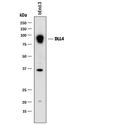
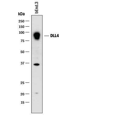
- Detection of Mouse DLL4 by Western Blot. Western blot shows lysates of bEnd.3 mouse endothelioma cell line. PVDF membrane was probed with 2 µg/mL of Goat Anti-Mouse DLL4 Antigen Affinity-purified Polyclonal Antibody (Catalog # AF1389) followed by HRP-conjugated Anti-Goat IgG Secondary Antibody (Catalog # HAF017). A specific band was detected for DLL4 at approximately 90 kDa (as indicated). This experiment was conducted under reducing conditions and using Immunoblot Buffer Group 1.
Supportive validation
- Submitted by
- R&D Systems (provider)
- Main image
- Experimental details
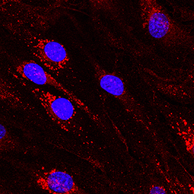
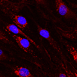
- DLL4 in bEnd.3 Mouse Cell Line. DLL4 was detected in immersion fixed bEnd.3 mouse endothelioma cell line using Goat Anti-Mouse DLL4 Antigen Affinity-purified Polyclonal Antibody (Catalog # AF1389) at 10 µg/mL for 3 hours at room temperature. Cells were stained using the NorthernLights™ 557-conjugated Anti-Goat IgG Secondary Antibody (red; Catalog # NL001) and counterstained with DAPI (blue). Specific staining was localized to cytoplasm. View our protocol for Fluorescent ICC Staining of Cells on Coverslips.