Explore
 Validate
 Learn
Learn Western blot
 ELISA
ELISAAntibody data
- Antibody Data
- Antigen structure
- References [0]
- Comments [0]
- Validations
- ELISA [2]
- Immunohistochemistry [1]
Submit
Validation data
Reference
Comment
Report error
- Product number
- LS-C745290 - Provider product page

- Provider
- LSBio
- Product name
- DLL4 Antibody (Internal) LS-C745290
- Antibody type
- Polyclonal
- Description
- Affinity purified
- Reactivity
- Human
- Host
- Rabbit
- Isotype
- IgG
- Storage
- Store vial at -20°C or below prior to opening. Dilute 1:10 to minimize loss. Store the vial at -20°C or below after dilution. Avoid freeze-thaw cycles.
No comments: Submit comment
Supportive validation
- Submitted by
- LSBio (provider)
- Enhanced method
- Genetic validation
- Main image

- Experimental details
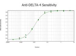
- ELISA results of purified Rabbit anti-DELTA-4 Antibody tested against BSA-conjugated peptide of immunizing peptide. Each well was coated in duplicate with 0.1µg of conjugate. The starting dilution of antibody was 5µg/ml and the X-axis represents the Log10 of a 3-fold dilution. This titration is a 4-parameter curve fit where the IC50 is defined as the titer of the antibody. Assay performed using 3% fish gel, Goat anti-Rabbit IgG Antibody Peroxidase Conjugated (Min X Bv Ch Gt GP Ham Hs Hu Ms Rt & Sh Serum Proteins)

- Submitted by
- LSBio (provider)
- Main image
- Experimental details
- ELISA results of purified Rabbit anti-DELTA-4 Antibody tested against BSA-conjugated peptide of immunizing peptide. Each well was coated in duplicate with 0.1µg of conjugate. The starting dilution of antibody was 5µg/ml and the X-axis represents the Log10 of a 3-fold dilution. This titration is a 4-parameter curve fit where the IC50 is defined as the titer of the antibody. Assay performed using 3% fish gel, Goat anti-Rabbit IgG Antibody Peroxidase Conjugated (Min X Bv Ch Gt GP Ham Hs Hu Ms Rt & Sh Serum Proteins)

Supportive validation
- Submitted by
- LSBio (provider)
- Main image
- Experimental details
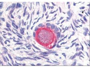
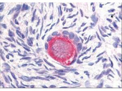
- Affinity Purified anti-Delta-4 antibody was used at 20 µg/ml to detect Delta-4 in a variety of tissues including colon, liver, skeletal muscle, ovary, pancreas, prostate, testes, thymus, tonsil and uterus. In contrast to reported findings, no staining was observed in vascular tissue. This image shows Delta-4 staining of human ovary. Tissue was formalin-fixed and paraffin embedded.