Explore
 Validate
 Learn
Learn Western blot
 Immunohistochemistry
Antibody data
- Antibody Data
- Antigen structure
- References [7]
- Comments [0]
- Validations
- Immunohistochemistry [1]
Submit
Validation data
Reference
Comment
Report error
- Product number
- MAB1389 - Provider product page

- Provider
- R&D Systems
- Product name
- Human/Mouse DLL4 Antibody
- Antibody type
- Monoclonal
- Description
- Protein A or G purified from hybridoma culture supernatant. Detects mouse DLL4 in direct ELISAs and Western blots. In direct ELISAs and Western blots, this antibody shows 100% cross-reactivity with recombinant human DLL4.
- Reactivity
- Human, Mouse
- Host
- Rat
- Conjugate
- Unconjugated
- Antigen sequence
Q9JI71- Isotype
- IgG
- Antibody clone number
- 207822
- Vial size
- 500 ug
- Concentration
- LYOPH
- Storage
- Use a manual defrost freezer and avoid repeated freeze-thaw cycles. 12 months from date of receipt, -20 to -70 °C as supplied. 1 month, 2 to 8 °C under sterile conditions after reconstitution. 6 months, -20 to -70 °C under sterile conditions after reconstitution.
Submitted references Muscle Satellite Cell Cross-Talk with a Vascular Niche Maintains Quiescence via VEGF and Notch Signaling.
MEF2 transcription factors are key regulators of sprouting angiogenesis.
Suppression of ischemia in arterial occlusive disease by JNK-promoted native collateral artery development.
Quantitative assessment of angiogenesis, perfused blood vessels and endothelial tip cells in the postnatal mouse brain.
The alarmin IL-33 is a notch target in quiescent endothelial cells.
KSHV-induced notch components render endothelial and mural cell characteristics and cell survival.
CD8- DCs induce IL-12-independent Th1 differentiation through Delta 4 Notch-like ligand in response to bacterial LPS.
Verma M, Asakura Y, Murakonda BSR, Pengo T, Latroche C, Chazaud B, McLoon LK, Asakura A
Cell stem cell 2018 Oct 4;23(4):530-543.e9
Cell stem cell 2018 Oct 4;23(4):530-543.e9
MEF2 transcription factors are key regulators of sprouting angiogenesis.
Sacilotto N, Chouliaras KM, Nikitenko LL, Lu YW, Fritzsche M, Wallace MD, Nornes S, García-Moreno F, Payne S, Bridges E, Liu K, Biggs D, Ratnayaka I, Herbert SP, Molnár Z, Harris AL, Davies B, Bond GL, Bou-Gharios G, Schwarz JJ, De Val S
Genes & development 2016 Oct 15;30(20):2297-2309
Genes & development 2016 Oct 15;30(20):2297-2309
Suppression of ischemia in arterial occlusive disease by JNK-promoted native collateral artery development.
Ramo K, Sugamura K, Craige S, Keaney JF, Davis RJ
eLife 2016 Aug 9;5
eLife 2016 Aug 9;5
Quantitative assessment of angiogenesis, perfused blood vessels and endothelial tip cells in the postnatal mouse brain.
Wälchli T, Mateos JM, Weinman O, Babic D, Regli L, Hoerstrup SP, Gerhardt H, Schwab ME, Vogel J
Nature protocols 2015 Jan;10(1):53-74
Nature protocols 2015 Jan;10(1):53-74
The alarmin IL-33 is a notch target in quiescent endothelial cells.
Sundlisaeter E, Edelmann RJ, Hol J, Sponheim J, Küchler AM, Weiss M, Udalova IA, Midwood KS, Kasprzycka M, Haraldsen G
The American journal of pathology 2012 Sep;181(3):1099-111
The American journal of pathology 2012 Sep;181(3):1099-111
KSHV-induced notch components render endothelial and mural cell characteristics and cell survival.
Liu R, Li X, Tulpule A, Zhou Y, Scehnet JS, Zhang S, Lee JS, Chaudhary PM, Jung J, Gill PS
Blood 2010 Jan 28;115(4):887-95
Blood 2010 Jan 28;115(4):887-95
CD8- DCs induce IL-12-independent Th1 differentiation through Delta 4 Notch-like ligand in response to bacterial LPS.
Skokos D, Nussenzweig MC
The Journal of experimental medicine 2007 Jul 9;204(7):1525-31
The Journal of experimental medicine 2007 Jul 9;204(7):1525-31
No comments: Submit comment
Supportive validation
- Submitted by
- R&D Systems (provider)
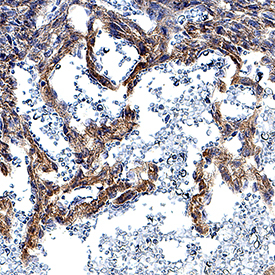
- Main image
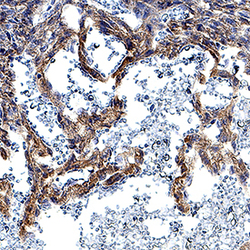
- Experimental details
- DLL4 in Mouse Embryonic Heart. DLL4 was detected in immersion fixed frozen sections of mouse embryonic heart using Rat Anti-Human/Mouse DLL4 Monoclonal Antibody (Catalog # MAB1389) at 8 µg/mL overnight at 4 °C. Tissue was stained using the Anti-Rat HRP-DAB Cell & Tissue Staining Kit (brown; Catalog # CTS017) and counterstained with hematoxylin (blue). Specific staining was localized to developing cardiomyocytes. View our protocol for Chromogenic IHC Staining of Frozen Tissue Sections.