 Explore
 Validate
 Learn
Learn Western blot
 Immunohistochemistry
Antibody data
- Antibody Data
- Antigen structure
- References [9]
- Comments [0]
- Validations
- Western blot [1]
- Immunohistochemistry [1]
Submit
Validation data
Reference
Comment
Report error
- Product number
- HPA000798 - Provider product page

- Provider
- Atlas Antibodies
- Proper citation
- Atlas Antibodies Cat#HPA000798, RRID:AB_1079090
- Product name
- Anti-HSPA2
- Antibody type
- Polyclonal
- Description
- Polyclonal Antibody against Human HSPA2, Gene description: heat shock 70kDa protein 2, Validated applications: WB, IHC, Uniprot ID: P54652, Storage: Store at +4°C for short term storage. Long time storage is recommended at -20°C.
- Reactivity
- Human, Mouse, Rat
- Host
- Rabbit
- Conjugate
- Unconjugated
- Isotype
- IgG
- Vial size
- 100 µl
- Concentration
- 0.1 mg/ml
- Storage
- Store at +4°C for short term storage. Long time storage is recommended at -20°C.
- Handling
- The antibody solution should be gently mixed before use.
Submitted references Molecular Chaperone HSPA2 Distribution During Hyaluronic Acid Selection in Human Sperm
4‐phenylbutyric acid—Identity crisis; can it act as a translation inhibitor?
Multi-omics integration identifies key upstream regulators of pathomechanisms in hypertrophic cardiomyopathy due to truncating MYBPC3 mutations
Transcriptomic analysis identifies a tumor subtype mRNA classifier for invasive non-functioning pituitary neuroendocrine tumor diagnostics
Translocation of molecular chaperones to the titin springs is common in skeletal myopathy patients and affects sarcomere function
Expression, function, and regulation of the testis-enriched heat shock HSPA2 gene in rodents and humans
HSP70-binding protein HSPBP1 regulates chaperone expression at a posttranslational level and is essential for spermatogenesis.
The molecular chaperone Hsp90α is required for meiotic progression of spermatocytes beyond pachytene in the mouse.
Systematically generated antibodies against human gene products: High throughput screening on sections from the rat nervous system
Gómez-Torres M, Huerta-Retamal N, Sáez-Espinosa P, Robles-Gómez L, Avilés M, Aizpurua J
Reproductive Sciences 2022;30(4):1176-1185
Reproductive Sciences 2022;30(4):1176-1185
4‐phenylbutyric acid—Identity crisis; can it act as a translation inhibitor?
Stein D, Slobodnik Z, Tam B, Einav M, Akabayov B, Berstein S, Toiber D
Aging Cell 2022;21(12)
Aging Cell 2022;21(12)
Multi-omics integration identifies key upstream regulators of pathomechanisms in hypertrophic cardiomyopathy due to truncating MYBPC3 mutations
Pei J, Schuldt M, Nagyova E, Gu Z, el Bouhaddani S, Yiangou L, Jansen M, Calis J, Dorsch L, Blok C, van den Dungen N, Lansu N, Boukens B, Efimov I, Michels M, Verhaar M, de Weger R, Vink A, van Steenbeek F, Baas A, Davis R, Uh H, Kuster D, Cheng C, Mokry M, van der Velden J, Asselbergs F, Harakalova M
Clinical Epigenetics 2021;13(1)
Clinical Epigenetics 2021;13(1)
Transcriptomic analysis identifies a tumor subtype mRNA classifier for invasive non-functioning pituitary neuroendocrine tumor diagnostics
Bao X, Wang G, Yu S, Sun J, He L, Zhao H, Ma Y, Wang F, Wang X, Wang R, Yu J
Theranostics 2021;11(1):132-146
Theranostics 2021;11(1):132-146
Translocation of molecular chaperones to the titin springs is common in skeletal myopathy patients and affects sarcomere function
Unger A, Beckendorf L, Böhme P, Kley R, von Frieling-Salewsky M, Lochmüller H, Schröder R, Fürst D, Vorgerd M, Linke W
Acta Neuropathologica Communications 2017;5(1)
Acta Neuropathologica Communications 2017;5(1)
Expression, function, and regulation of the testis-enriched heat shock HSPA2 gene in rodents and humans
Scieglinska D, Krawczyk Z
Cell Stress and Chaperones 2015;20(2):221-235
Cell Stress and Chaperones 2015;20(2):221-235
HSP70-binding protein HSPBP1 regulates chaperone expression at a posttranslational level and is essential for spermatogenesis.
Rogon C, Ulbricht A, Hesse M, Alberti S, Vijayaraj P, Best D, Adams IR, Magin TM, Fleischmann BK, Höhfeld J
Molecular biology of the cell 2014 Aug 1;25(15):2260-71
Molecular biology of the cell 2014 Aug 1;25(15):2260-71
The molecular chaperone Hsp90α is required for meiotic progression of spermatocytes beyond pachytene in the mouse.
Grad I, Cederroth CR, Walicki J, Grey C, Barluenga S, Winssinger N, De Massy B, Nef S, Picard D
PloS one 2010 Dec 31;5(12):e15770
PloS one 2010 Dec 31;5(12):e15770
Systematically generated antibodies against human gene products: High throughput screening on sections from the rat nervous system
Mulder J, Wernérus H, Shi T, Pontén F, Hober S, Uhlén M, Hökfelt T
Neuroscience 2007;146(4):1689-1703
Neuroscience 2007;146(4):1689-1703
No comments: Submit comment
Enhanced validation
- Submitted by
- Atlas Antibodies (provider)
- Enhanced method
- Genetic validation
- Main image
- Experimental details
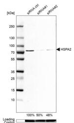
- Western blot analysis in A-549 cells transfected with control siRNA, target specific siRNA probe #1 and #2, using Anti-HSPA2 antibody. Remaining relative intensity is presented. Loading control: Anti-GAPDH.
- Sample type
- Human
- Protocol
- Protocol

Supportive validation
- Submitted by
- Atlas Antibodies (provider)
- Enhanced method
- Orthogonal validation
- Main image
- Experimental details
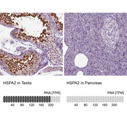
- Immunohistochemistry analysis in human testis and pancreas tissues using HPA000798 antibody. Corresponding HSPA2 RNA-seq data are presented for the same tissues.
- Sample type
- Human
- Protocol
- Protocol
