Explore
 Validate
 Learn
LearnPB9237
antibody from Boster Biological Technology
Targeting: HSPB1
CMT2F, Hs.76067, Hsp25, HSP27, HSP28
 Western blot
 Immunocytochemistry
Antibody data
- Antibody Data
- Antigen structure
- References [1]
- Comments [0]
- Validations
- Western blot [1]
Submit
Validation data
Reference
Comment
Report error
- Product number
- PB9237 - Provider product page

- Provider
- Boster Biological Technology
- Product name
- Anti-Hsp27/HSPB1 Antibody Picoband™
- Antibody type
- Polyclonal
- Description
- Polyclonal antibody for HSP27/HSPB1 detection. Host: Rabbit.Size: 100μg/vial. Tested applications: WB, IHC-P, IHC-F, ICC/IF, FCM. Reactive species: Human. HSP27/HSPB1 information: Molecular Weight: 22783 MW; Subcellular Localization: Cytoplasm. Nucleus. Cytoplasm, cytoskeleton, spindle. Cytoplasmic in interphase cells. Colocalizes with mitotic spindles in mitotic cells. Translocates to the nucleus during heat shock and resides in sub-nuclear structures known as SC35 speckles or nuclear splicing speckles; Tissue Specificity: Detected in all tissues tested: skeletal muscle, heart, aorta, large intestine, small intestine, stomach, esophagus, bladder, adrenal gland, thyroid, pancreas, testis, adipose tissue, kidney, liver, spleen, cerebral cortex, blood serum and cerebrospinal fluid. Highest levels are found in the heart and in tissues composed of striated and smooth muscle.
- Reactivity
- Human
- Host
- Rabbit
- Vial size
- 100μg/vial
- Concentration
- Add 0.2ml of distilled water will yield a concentration of 500ug/ml.
- Storage
- At -20°C for one year. After reconstitution, at 4°C for one month. It can also be aliquoted and stored frozen at -20°C for a longer time. Avoid repeated freezing and thawing.
- Handling
- Add 0.2ml of distilled water will yield a concentration of 500ug/ml.
Submitted references LRRC4 inhibits the proliferation of human glioma cells by modulating the expression of STMN1 and microtubule polymerization.
Wang R, Wang Z, Yang J, Liu X, Wang L, Guo X, Zeng F, Wu M, Li G
Journal of cellular biochemistry 2011 Dec;112(12):3621-9
Journal of cellular biochemistry 2011 Dec;112(12):3621-9
No comments: Submit comment
Supportive validation
- Submitted by
- Boster Biological Technology (provider)
- Main image
- Experimental details
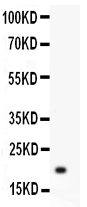
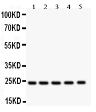
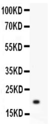
- Western blot analysis of HSP27 using anti-HSP27 antibody (PB9237). Electrophoresis was performed on a 5-20% SDS-PAGE gel at 70V (Stacking gel) / 90V (Resolving gel) for 2-3 hours. Lane 1: Recombinant Human HSP27 Protein 0.5ng After Electrophoresis, proteins were transferred to a Nitrocellulose membrane at 150mA for 50-90 minutes. Blocked the membrane with 5% Non-fat Milk/ TBS for 1.5 hour at RT. The membrane was incubated with rabbit anti-HSP27 antigen affinity purified polyclonal antibody (Catalog # PB9237) at 0.5 μg/mL overnight at 4°C, then washed with TBS-0.1%Tween 3 times with 5 minutes each and probed with a goat anti-rabbit IgG-HRP secondary antibody at a dilution of 1:10000 for 1.5 hour at RT. The signal is developed using an Enhanced Chemiluminescent detection (ECL) kit (Catalog # EK1002) with Tanon 5200 system. A specific band was detected for HSP27 at approximately 19KD. The expected band size for HSP27 is at 19KD.
- Additional image