 Explore
 Validate
 Learn
Learn Western blot
Antibody data
- Antibody Data
- Antigen structure
- References [3]
- Comments [0]
- Validations
- Western blot [2]
Submit
Validation data
Reference
Comment
Report error
- Product number
- AF1580 - Provider product page

- Provider
- Novus Biologicals
- Product name
- Rabbit Polyclonal HSP27 Antibody
- Antibody type
- Polyclonal
- Description
- Antigen Affinity-purified. Detects recombinant and endogenous human and mouse HSP27 in Western blots.
- Reactivity
- Human, Mouse
- Host
- Rabbit
- Conjugate
- Unconjugated
- Isotype
- IgG
- Vial size
- 50 ug
- Concentration
- LYOPH
- Storage
- Use a manual defrost freezer and avoid repeated freeze-thaw cycles. 12 months from date of receipt, -20 to -70 degreesC as supplied. 1 month, 2 to 8 degreesC under sterile conditions after reconstitution. 6 months, -20 to -70 degreesC under sterile conditions after reconstitution.
Submitted references Heat shock protein 27 differentiates tolerogenic macrophages that may support human breast cancer progression.
The heat shock response and chaperones/heat shock proteins in brain tumors: surface expression, release, and possible immune consequences.
Phosphatidylinositol 3-kinase/Akt plays a role in sphingosine 1-phosphate-stimulated HSP27 induction in osteoblasts.
Banerjee S, Lin CF, Skinner KA, Schiffhauer LM, Peacock J, Hicks DG, Redmond EM, Morrow D, Huston A, Shayne M, Langstein HN, Miller-Graziano CL, Strickland J, O'Donoghue L, De AK
Cancer research 2011 Jan 15;71(2):318-27
Cancer research 2011 Jan 15;71(2):318-27
The heat shock response and chaperones/heat shock proteins in brain tumors: surface expression, release, and possible immune consequences.
Graner MW, Cumming RI, Bigner DD
The Journal of neuroscience : the official journal of the Society for Neuroscience 2007 Oct 17;27(42):11214-27
The Journal of neuroscience : the official journal of the Society for Neuroscience 2007 Oct 17;27(42):11214-27
Phosphatidylinositol 3-kinase/Akt plays a role in sphingosine 1-phosphate-stimulated HSP27 induction in osteoblasts.
Takai S, Tokuda H, Matsushima-Nishiwaki R, Hanai Y, Kato K, Kozawa O
Journal of cellular biochemistry 2006 Aug 1;98(5):1249-56
Journal of cellular biochemistry 2006 Aug 1;98(5):1249-56
No comments: Submit comment
Supportive validation
- Submitted by
- Novus Biologicals (provider)
- Main image

- Experimental details
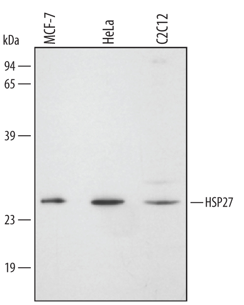
- Detection of Human/Mouse HSP27 by Western Blot. Western blot shows lysates of MCF-7 human breast cancer cell line, HeLa human cervical epithelial carcinoma cell line, and C2C12 mouse myoblast cell line. PVDF membrane was probed with 0.1 µg/mL of Rabbit Anti-Human/Mouse HSP27 Antigen Affinity-purified Polyclonal Antibody (Catalog # AF1580) followed by HRP-conjugated Anti-Rabbit IgG Secondary Antibody (Catalog # HAF008). A specific band was detected for HSP27 at approximately 27 kDa (as indicated). This experiment was conducted under reducing conditions and using Immunoblot Buffer Group 2.
- Submitted by
- Novus Biologicals (provider)
- Main image

- Experimental details
- Detection of Mouse HSP27 by Simple WesternTM. Simple Western lane view shows lysates of C2C12 mouse myoblast cell line, loaded at 0.2 mg/mL. A specific band was detected for HSP27 at approximately 31 kDa (as indicated) using 0.5 µg/mL of Rabbit Anti-Human/Mouse HSP27 Antigen Affinity-purified Polyclonal Antibody (Catalog # AF1580). This experiment was conducted under reducing conditions and using the 12-230 kDa separation system.
