 Explore
 Validate
 Learn
Learn Western blot
 Immunohistochemistry
Antibody data
- Antibody Data
- Antigen structure
- References [0]
- Comments [0]
- Validations
- Western blot [1]
- Immunocytochemistry [1]
- Immunoprecipitation [1]
Submit
Validation data
Reference
Comment
Report error
- Product number
- GTX25581 - Provider product page

- Provider
- GeneTex
- Proper citation
- GeneTex Cat#GTX25581, RRID:AB_371723
- Product name
- HSP27 (phospho Ser15) antibody
- Antibody type
- Polyclonal
- Reactivity
- Human, Rat, Rabbit
- Host
- Rabbit
No comments: Submit comment
Supportive validation
- Submitted by
- GeneTex (provider)
- Main image
- Experimental details
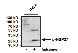
- Western blot analysis of Phospho Heat Shock Protein 27 (p-HSP27) pSer15 was performed by loading 50ug of HeLa cell lysates from untreated cells (left lane) or cells treated with 10uM Anisomycin (right lane) for 30 minutes and 15ul onto a 4-20% Tris-HCl polyacrylamide gel. Proteins were transferred to a PVDF membrane and blocked with 5% BSA/TBST for at least 1 hour. The membrane was probed with a p-HSP27 pSer15 polyclonal antibody at a dilution of 1:500 overnight at 4¢XC on a rocking platform, washed with TBS-0.1%Tween-20, and probed with a goat anti-rabbit IgG-HRP secondary antibody at a dilution of 1:20,000 for at least 1 hour. Chemiluminescent detection was performed.

Supportive validation
- Submitted by
- GeneTex (provider)
- Main image

- Experimental details
- Immunofluorescent analysis of Phospho Heat Shock Protein 27 (p-HSP27) pSer15 (green) in HeLa cells either left untreated (left panel) or treated with 10uM Anisomysin (right panel) for 30 minutes. Formalin fixed cells were permeabilized with 0.1% Triton X-100 in TBS for 10 minutes at room temperature and blocked with 1% Blocker BSA for 15 minutes at room temperature. Cells were probed with a p-HSP27 pSer15 polyclonal antibody at a dilution of 1:50 for at least 1 hour at room temperature, washed with PBS, and incubated with DyLight 488 goat anti-rabbit IgG secondary antibody at a dilution of 1:400 for 30 minutes at room temperature. F-Actin (red) was stained with DyLight 554 Phalloidin and nuclei (blue) were stained with Hoechst dye.

Supportive validation
- Submitted by
- GeneTex (provider)
- Main image

- Experimental details
- Immunoprecipitation of Phospho Heat Shock Protein 27 (p-HSP27) pSer15 was performed on HeLa cells treated with 10uM Anisomysin for 30 minutes. Antigen-antibody complexes were formed by incubating 500ug of whole cell lysate with 3ug of p-HSP27 pSer15 polyclonal antibody (GTX25581) overnight on a rocking platform at 4¢XC. The immune complexes were captured on 50ul Protein A/G Agarose, washed extensively, and eluted. Samples were then resolved on a 4-20% Tris-HCl polyacrylamide gel, transferred to a PVDF membrane, and blocked with 5% BSA/TBST for at least 1 hour. The membrane was probed with a p-HSP27 pSer15 polyclonal antibody (GTX25581) at a dilution of 1:500 overnight rotating at 4¢XC, washed with TBST, and probed with an IP detection reagent at a dilution of 1:1000 for at least 1 hour. Chemiluminescent detection was performed
