Explore
 Validate
 Learn
Learn14-9112-80
antibody from Invitrogen Antibodies
Targeting: HSPB1
CMT2F, Hs.76067, Hsp25, HSP27, HSP28
 Western blot
 ELISA
ELISA Immunocytochemistry
Antibody data
- Antibody Data
- Antigen structure
- References [4]
- Comments [0]
- Validations
- Immunocytochemistry [4]
- Immunohistochemistry [1]
- Other assay [2]
Submit
Validation data
Reference
Comment
Report error
- Product number
- 14-9112-80 - Provider product page

- Provider
- Invitrogen Antibodies
- Product name
- HSP27 Monoclonal Antibody (STRSN), eBioscience™
- Antibody type
- Monoclonal
- Antigen
- Other
- Description
- Description: The monoclonal antibody STRSN recognizes human heat shock protein 27 (HSP27). The heat shock proteins are a large family of proteins that are induced in response to stressors such as changing temperatures, cytokine levels, and presence of various chemicals. Heat shock proteins serve as molecular chaperones, binding to other proteins to maintain proper protein structure or by folding denatured proteins and preventing activation of caspases that could lead to apoptosis. Unlike the larger HSP proteins, HSP27 belongs to a unique subgroup that is involved in apoptosis regulation. Expression of HSP27 is constitutive, but upon stress HSP27 is upregulated and can relocate from the cytoplasm to the nucleus. Increased HSP27 expression with certain carcinomas has been observed. In breast cancer, HSP27 expression has been shown to be responsive to estrogen levels. In both breast cancer and melanoma, high levels of HSP27 correlate with more aggressive tumors and decreased survival. Increased levels of HSP27 in the serum of patients with Chronic Obstructive Pulmonary Disease (COPD), breast cancer and melanoma may serve as a potential diagnostic marker. No crossreactivity is observed with HSP70, 90, 104 or 110. Applications Reported: This STRSN antibody has been reported for use in western blotting, immunohistochemical staining of formalin-fixed paraffin embedded tissue sections, ELISA, and immunocytochemistry. Applications Tested: This STRSN antibody has been tested by western blot of reduced HeLa cell lysate, immunocytochemistry on fixed and permeabilized A549 cells, and by immunohistochemistry on formalin-fixed paraffin embedded (FFPE) human tissue with either low or high pH antigen retreival at less than or equal to 1 µg/mL. It is recommended that the antibody be carefully titrated for optimal performance in the assay of interest. Purity: Greater than 90%, as determined by SDS-PAGE. Aggregation: Less than 10%, as determined by HPLC. Filtration: 0.2 µm post-manufacturing filtered.
- Reactivity
- Human
- Host
- Mouse
- Isotype
- IgG
- Antibody clone number
- STRSN
- Vial size
- 25 μg
- Concentration
- 0.5 mg/mL
- Storage
- 4°C
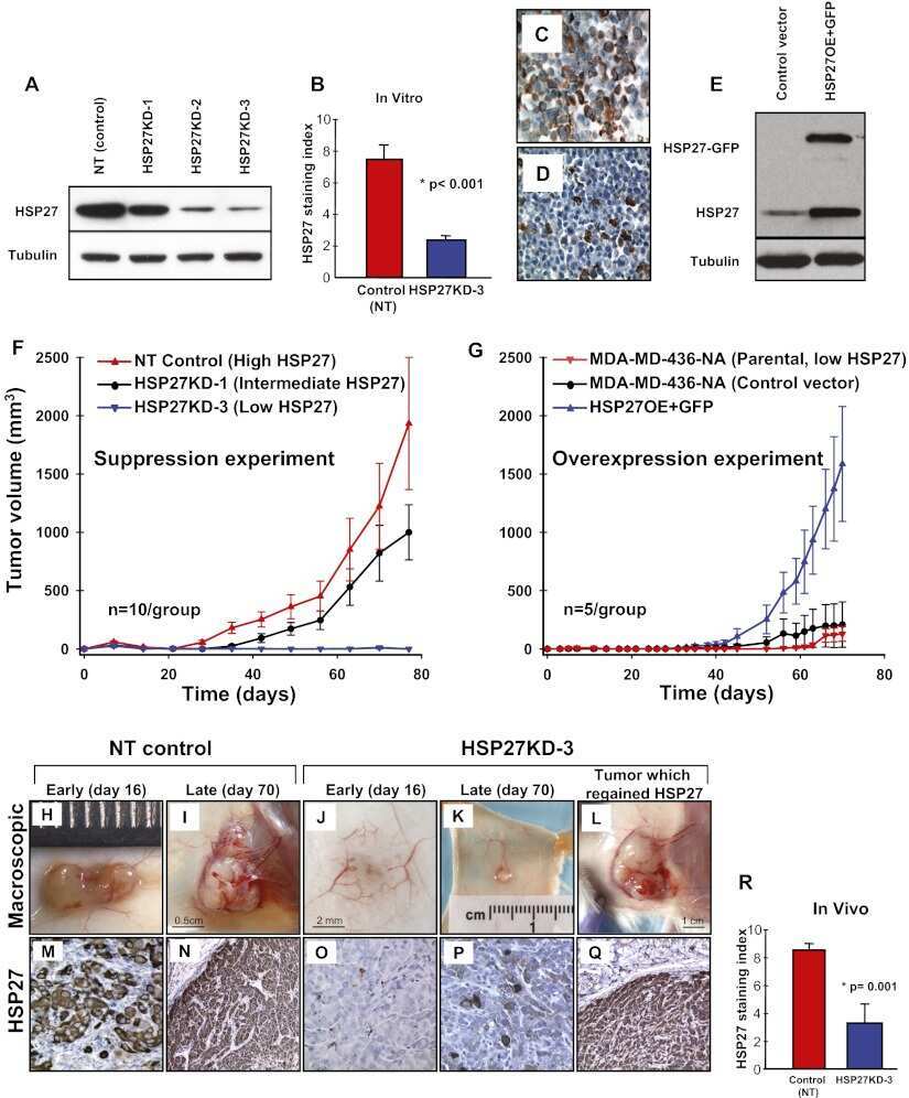
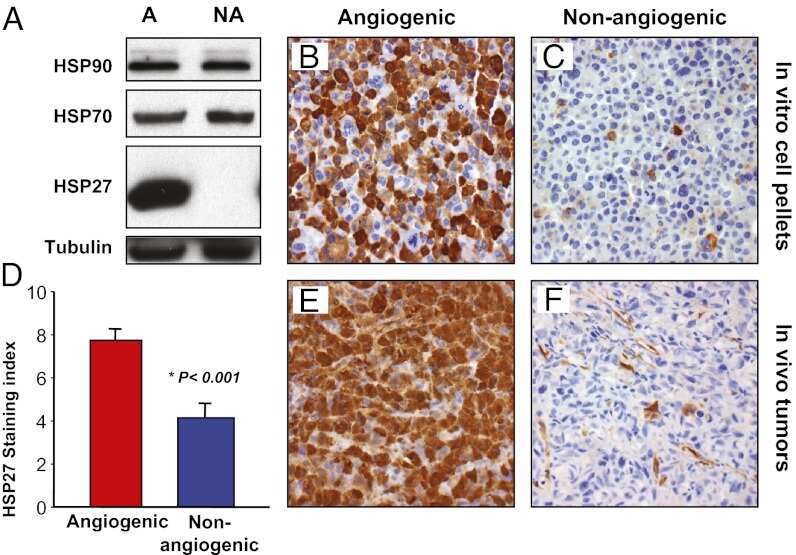
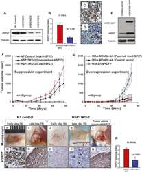
Submitted references Suppression of heat shock protein 27 induces long-term dormancy in human breast cancer.
The small heat shock protein 27 is a key regulator of CD8+ CD57+ lymphocyte survival.
Roles of heat-shock proteins in innate and adaptive immunity.
Human HSP27 is phosphorylated at serines 78 and 82 by heat shock and mitogen-activated kinases that recognize the same amino acid motif as S6 kinase II.
Straume O, Shimamura T, Lampa MJ, Carretero J, Øyan AM, Jia D, Borgman CL, Soucheray M, Downing SR, Short SM, Kang SY, Wang S, Chen L, Collett K, Bachmann I, Wong KK, Shapiro GI, Kalland KH, Folkman J, Watnick RS, Akslen LA, Naumov GN
Proceedings of the National Academy of Sciences of the United States of America 2012 May 29;109(22):8699-704
Proceedings of the National Academy of Sciences of the United States of America 2012 May 29;109(22):8699-704
The small heat shock protein 27 is a key regulator of CD8+ CD57+ lymphocyte survival.
Wood KL, Voss OH, Huang Q, Parihar A, Mehta N, Batra S, Doseff AI
Journal of immunology (Baltimore, Md. : 1950) 2010 May 15;184(10):5582-8
Journal of immunology (Baltimore, Md. : 1950) 2010 May 15;184(10):5582-8
Roles of heat-shock proteins in innate and adaptive immunity.
Srivastava P
Nature reviews. Immunology 2002 Mar;2(3):185-94
Nature reviews. Immunology 2002 Mar;2(3):185-94
Human HSP27 is phosphorylated at serines 78 and 82 by heat shock and mitogen-activated kinases that recognize the same amino acid motif as S6 kinase II.
Landry J, Lambert H, Zhou M, Lavoie JN, Hickey E, Weber LA, Anderson CW
The Journal of biological chemistry 1992 Jan 15;267(2):794-803
The Journal of biological chemistry 1992 Jan 15;267(2):794-803
No comments: Submit comment
Supportive validation
- Submitted by
- Invitrogen Antibodies (provider)
- Main image

- Experimental details
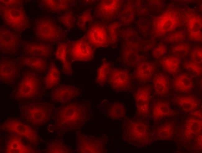
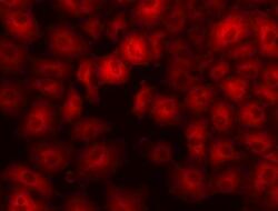
- Immunocytochemistry of fixed and permeabilized A549 cells using 1 µg/mL of Anti-Human Heat Shock Protein 27 Purified followed by Anti-Mouse IgG TRITC.
- Submitted by
- Invitrogen Antibodies (provider)
- Main image
- Experimental details
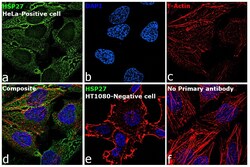
- Immunofluorescence analysis of HSP27 was performed using 70% confluent log phase HeLa cells. The cells were fixed with 4% paraformaldehyde for 10 minutes, permeabilized with 0.1% Triton™ X-100 for 15 minutes, and blocked with 2% BSA for 45 minutes at room temperature. The cells were labeled with HSP27 Monoclonal Antibody (STRSN), eBioscience™ (Product # 14-9112-80) at 2.5 µg/mL in 0.1% BSA, incubated at 4 degree celsius overnight and then labeled with Goat anti-Mouse IgG (H+L) Highly Cross-Adsorbed Secondary Antibody, Alexa Fluor Plus 488 (Product # A32723), (1:2000), for 45 minutes at room temperature (Panel a: Green). Nuclei (Panel b: Blue) were stained with ProLong™ Diamond Antifade Mountant with DAPI (Product # P36962). F-actin (Panel c: Red) was stained with Rhodamine Phalloidin (Product # R415, 1:300). Panel d represents the merged image showing cytoplasmic localization. Panel e represents negative cell line (HT-1080) with no signal. Panel f represents control cells with no primary antibody to assess background. The images were captured at 60X magnification.

- Submitted by
- Invitrogen Antibodies (provider)
- Main image

- Experimental details
- Immunofluorescence analysis of HSP27 was performed using 70% confluent log phase HeLa cells. The cells were fixed with 4% paraformaldehyde for 10 minutes, permeabilized with 0.1% Triton™ X-100 for 15 minutes, and blocked with 2% BSA for 45 minutes at room temperature. The cells were labeled with HSP27 Monoclonal Antibody (STRSN), eBioscience™ (Product # 14-9112-80) at 2.5 µg/mL in 0.1% BSA, incubated at 4 degree celsius overnight and then labeled with Goat anti-Mouse IgG (H+L) Highly Cross-Adsorbed Secondary Antibody, Alexa Fluor Plus 488 (Product # A32723), (1:2000), for 45 minutes at room temperature (Panel a: Green). Nuclei (Panel b: Blue) were stained with ProLong™ Diamond Antifade Mountant with DAPI (Product # P36962). F-actin (Panel c: Red) was stained with Rhodamine Phalloidin (Product # R415, 1:300). Panel d represents the merged image showing cytoplasmic localization. Panel e represents negative cell line (HT-1080) with no signal. Panel f represents control cells with no primary antibody to assess background. The images were captured at 60X magnification.

- Submitted by
- Invitrogen Antibodies (provider)
- Main image
- Experimental details
- Immunocytochemistry of fixed and permeabilized A549 cells using 1 µg/mL of Anti-Human Heat Shock Protein 27 Purified followed by Anti-Mouse IgG TRITC.

Supportive validation
- Submitted by
- Invitrogen Antibodies (provider)
- Main image

- Experimental details
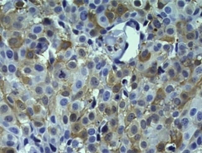
- Immunohistochemistry of formalin-fixed paraffin embedded human breast cancer using 1 µg/mL of Anti-Human Heat Shock Protein 27 Purified followed by Anti-Mouse IgG Biotin, Streptavidin HRP and DAB visualization.Nuclei are counterstained with hematoxylin.
Supportive validation
- Submitted by
- Invitrogen Antibodies (provider)
- Main image
- Experimental details
- NULL
- Submitted by
- Invitrogen Antibodies (provider)
- Main image
- Experimental details
- NULL