Explore
 Validate
 Learn
Learn Western blot
 Immunohistochemistry
Antibody data
- Antibody Data
- Antigen structure
- References [2]
- Comments [0]
- Validations
- Western blot [2]
- Immunocytochemistry [1]
- Immunoprecipitation [1]
- Flow cytometry [1]
Submit
Validation data
Reference
Comment
Report error
- Product number
- PA1-005 - Provider product page

- Provider
- Invitrogen Antibodies
- Product name
- Anti-Phospho-HSP27 (Ser85) Polyclonal Antibody
- Antibody type
- Polyclonal
- Antigen
- Synthetic peptide
- Description
- PA1-005 detects phosphorylated heat shock protein 27 (HSP27) from human and rat tissue samples. This antibody does not detect the unphosphorylated form of the protein. PA1-005 has been successfully used in Western blot, immunoprecipitation, and immunofluorescence procedures. By Western blot, this antibody detects an ~27 kDa protein representing phosphorylated HSP27(Ser85) from rat skeletal muscle extracts. The PA1-005 immunogen is a synthetic phosphopeptide corresponding to residues R(82) Q L S(p) S G V S E I R(92) C of rat HSP27. This sequence is completely conserved in mouse.
- Reactivity
- Human, Rat
- Host
- Rabbit
- Isotype
- IgG
- Vial size
- 100 µg
- Storage
- -20° C, Avoid Freeze/Thaw Cycles
Submitted references Increased cell surface Fas expression is necessary and sufficient to sensitize lung fibroblasts to Fas ligation-induced apoptosis: implications for fibroblast accumulation in idiopathic pulmonary fibrosis.
Neuroprotective preconditioning of rat brain cultures with ethanol: potential transduction by PKC isoforms and focal adhesion kinase upstream of increases in effector heat shock proteins.
Wynes MW, Edelman BL, Kostyk AG, Edwards MG, Coldren C, Groshong SD, Cosgrove GP, Redente EF, Bamberg A, Brown KK, Reisdorph N, Keith RC, Frankel SK, Riches DW
Journal of immunology (Baltimore, Md. : 1950) 2011 Jul 1;187(1):527-37
Journal of immunology (Baltimore, Md. : 1950) 2011 Jul 1;187(1):527-37
Neuroprotective preconditioning of rat brain cultures with ethanol: potential transduction by PKC isoforms and focal adhesion kinase upstream of increases in effector heat shock proteins.
Sivaswamy S, Neafsey EJ, Collins MA
The European journal of neuroscience 2010 Dec;32(11):1800-12
The European journal of neuroscience 2010 Dec;32(11):1800-12
No comments: Submit comment
Supportive validation
- Submitted by
- Invitrogen Antibodies (provider)
- Main image
- Experimental details
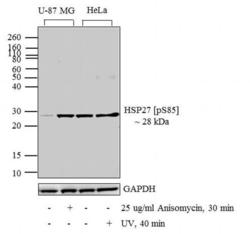
- Western blot analysis was performed on whole cell extracts (30 µg lysate) of U-87 MG (lane 1), U-87 MG treated for 30 minutes with 25 µg/mL of Anisomycin (lane 2), HeLa (lane 3) and HeLa treated with UV for 40 min (lane 4). The blots were probed with Anti-HSP27 (pS85) Rabbit Polyclonal Antibody (Product # PA1-005, 0.5-2 µg/mL) and detected by chemiluminescence using Goat anti-Rabbit IgG (H+L) Secondary Antibody, HRP conjugate (Product # G-21234, 1:5000 dilution). A 28 kDa band corresponding to HSP27 (pS85) was enhanced upon treatment. Known quantity of protein samples were electrophoresed using Novex® NuPAGE® 10 % Bis-Tris gel (Product # NP0301BOX), XCell SureLock™ Electrophoresis System (Product # EI0002) and Novex® Sharp Pre-Stained Protein Standard (Product # LC5800). Resolved proteins were then transferred onto a nitrocellulose membrane with PierceTM Power Blotter System (Product # 22834) The membrane was probed with the relevant primary and secondary Antibody following blocking with 5 % skimmed milk. Chemiluminescent detection was performed using Pierce™ ECL Western Blotting Substrate (Product # 32106).

- Submitted by
- Invitrogen Antibodies (provider)
- Main image
- Experimental details
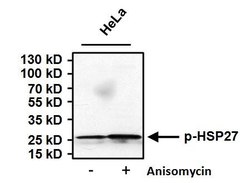
- Western blot analysis of Phospho Heat Shock Protein 27 (p-HSP27) pSer85 was performed by loading 50 µg of HeLa cell lysates from untreated cells (left lane) or cells treated with 10uM Anisomycin (right lane) for 30 minutes and 15 µL PageRuler Prestained Protein Ladder (Product # 26616) onto a 4-20% Tris-HCl polyacrylamide gel. Proteins were transferred to a PVDF membrane and blocked with 5% BSA/TBST for at least 1 hour. The membrane was probed with a p-HSP27 pSer85 polyclonal antibody (Product # PA1-005) at a concentration of 2 µg/mL overnight at 4°C on a rocking platform, washed in TBS-0.1%Tween-20, and probed with a goat anti-rabbit IgG-HRP secondary antibody (Product # 31460) at a dilution of 1:20,000 for at least 1 hour. Chemiluminescent detection was performed using SuperSignal West Pico (Product # 34080).
Supportive validation
- Submitted by
- Invitrogen Antibodies (provider)
- Main image
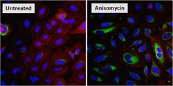
- Experimental details
- Immunofluorescent analysis of Phospho-HSP27 (p-HSP27) pSer85 (green) in HeLa cells either left untreated (left panel) or treated with 10uM Anisomysin (right panel) for 30 minutes. Formalin fixed cells were permeabilized with 0.1% Triton X-100 in TBS for 10 minutes at room temperature and blocked with 1% Blocker BSA (Product # 37525) for 15 minutes at room temperature. Cells were probed with a p-HSP27 pSer85 polyclonal antibody (Product # PA1-005), at a concentration of 20 µg/mL for at least 1 hour at room temperature, washed with PBS, and incubated with DyLight 488 goat anti-rabbit IgG secondary antibody (Product # 35552) at a dilution of 1:400 for 30 minutes at room temperature. F-Actin (red) was stained with DyLight 554 Phalloidin (Product # 21834) and nuclei (blue) were stained with Hoechst 33342 dye (Product # 62249). Images were taken on a Thermo Scientific ArrayScan at 20X magnification.

Supportive validation
- Submitted by
- Invitrogen Antibodies (provider)
- Main image
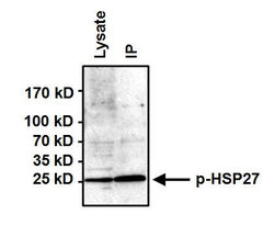
- Experimental details
- Immunoprecipitation of Phospho Heat Shock Protein (p-HSP27) pSer85 was performed on HeLa cells treated with 10uM Anisomysin for 30 minutes. Antigen-antibody complexes were formed by incubating 500µg of whole cell lysate with 3µg of a p-HSP27 pSer85 polyclonal antibody (Product # PA1-005) overnight on a rocking platform at 4¡C. The immune complexes were captured on 50µl Protein A/G Agarose (Product # 20421), washed extensively, and eluted with Lane Marker Reducing Sample Buffer (Product # 39000). HeLa cell lysate (50ug) was loaded as a positive control (left lane). Samples were resolved on a 4-20% Tris-HCl polyacrylamide gel, transferred to a PVDF membrane, and blocked with 5% BSA/TBST for at least 1 hour. The membrane was probed with a p-HSP27 pSer85 polyclonal antibody (Product # PA1-005) at a concentration of 2ug/ml overnight rotating at 4¡C, washed in TBST, and probed with Clean-Blot IP Detection Reagent (Product # 21230) at a dilution of 1:1000 for at least 1 hour. Chemiluminescent detection was performed using SuperSignal West Dura (Product # 34075).
Supportive validation
- Submitted by
- Invitrogen Antibodies (provider)
- Main image
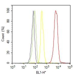
- Experimental details
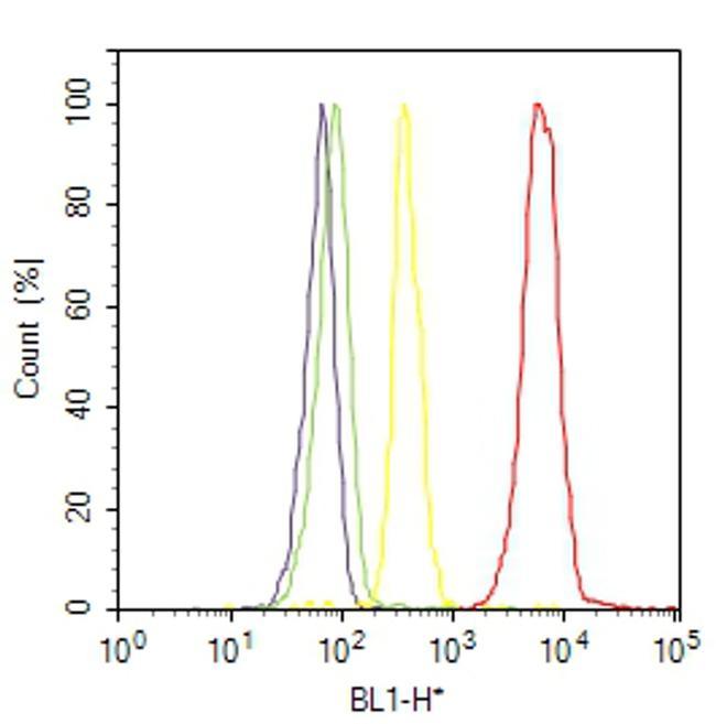
- Flow cytometry analysis of Phospho-HSP27 [pSer85] was done on HeLa cells treated with anisomycin (25ug/mL, 30 minutes). Cells were fixed with 70% ethanol for 10 minutes, permeabilized with 0.25% Triton™ X-100 for 20 minutes, and blocked with 5% BSA for 30 minutes at room temperature. Cells were labeled with Phospho-HSP27 [pSer85] Rabbit Polyclonal Antibody (PA1005, red histogram) or with rabbit isotype control (yellow histogram) at 3-5 ug/million cells in 2.5% BSA. After incubation at room temperature for 2 hours, the cells were labeled with Alexa Fluor® 488 Goat Anti-Rabbit Secondary Antibody (A11008) at a dilution of 1:400 for 30 minutes at room temperature. The representative 10,000 cells were acquired and analyzed for each sample using an Attune® Acoustic Focusing Cytometer. The purple histogram represents unstained control cells and the green histogram represents no-primary-antibody control.