Explore
 Validate
 Learn
Learn Western blot
Antibody data
- Antibody Data
- Antigen structure
- References [2]
- Comments [0]
- Validations
- Western blot [1]
- Immunocytochemistry [1]
Submit
Validation data
Reference
Comment
Report error
- Product number
- AF2314 - Provider product page

- Provider
- R&D Systems
- Product name
- Human/Mouse/Rat Phospho-HSP27 (S78/S82) Antibody
- Antibody type
- Polyclonal
- Description
- Antigen and protein A Affinity-purified. Detects human HSP27 when dually phosphorylated at S78/S82, and mouse and rat HSP27 phosphorylated at S86.
- Reactivity
- Human, Mouse, Rat
- Host
- Rabbit
- Conjugate
- Unconjugated
- Isotype
- IgG
- Vial size
- 50 ug
- Concentration
- LYOPH
- Storage
- Use a manual defrost freezer and avoid repeated freeze-thaw cycles. 12 months from date of receipt, -20 to -70 °C as supplied. 1 month, 2 to 8 °C under sterile conditions after reconstitution. 6 months, -20 to -70 °C under sterile conditions after reconstitution.
Submitted references A novel kinase function of a nucleoside-diphosphate-kinase homologue in Porphyromonas gingivalis is critical in subversion of host cell apoptosis by targeting heat-shock protein 27.
Survival of cancer stem cells under hypoxia and serum depletion via decrease in PP2A activity and activation of p38-MAPKAPK2-Hsp27.
Lee J, Roberts JS, Atanasova KR, Chowdhury N, Yilmaz Ö
Cellular microbiology 2018 May;20(5):e12825
Cellular microbiology 2018 May;20(5):e12825
Survival of cancer stem cells under hypoxia and serum depletion via decrease in PP2A activity and activation of p38-MAPKAPK2-Hsp27.
Lin SP, Lee YT, Wang JY, Miller SA, Chiou SH, Hung MC, Hung SC
PloS one 2012;7(11):e49605
PloS one 2012;7(11):e49605
No comments: Submit comment
Supportive validation
- Submitted by
- R&D Systems (provider)
- Main image
- Experimental details
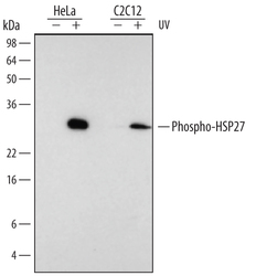
- Detection of Human and Mouse Phospho-HSP27 (S78/S82) by Western Blot. Western blot shows lysates of HeLa human cervical epithelial carcinoma cell line and C2C12 mouse myoblast cell line untreated (-) or treated (+) with 100 J/m2 UV-C for 30 minutes. PVDF membrane was probed with 0.1 µg/mL of Rabbit Anti-Human/Mouse/Rat Phospho-HSP27 (S78/S82) Antigen Affinity-purified Polyclonal Antibody (Catalog # AF2314), followed by HRP-conjugated Anti-Rabbit IgG Secondary Antibody (Catalog # HAF008). A specific band was detected for Phospho-HSP27 (S78/S82) at approximately 27 kDa (as indicated). This experiment was conducted under reducing conditions and using Immunoblot Buffer Group 1.

Supportive validation
- Submitted by
- R&D Systems (provider)
- Main image

- Experimental details
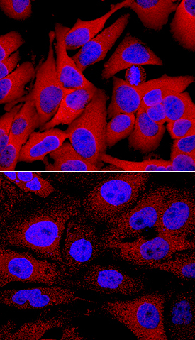
- Phospho-HSP27 (S78/S82) in HeLa Human Cell Line. HSP27 phosphorylated at S78/S82 was detected in immersion fixed HeLa human cervical epithelial carcinoma cell line unstimulated (lower panel) or stimulated with 20 mJ/cm2 ultraviolet radiation (upper panel) using Rabbit Anti-Human/Mouse/Rat Phospho-HSP27 (S78/S82) Antigen Affinity-purified Polyclonal Antibody (Catalog # AF2314) at 1 µg/mL for 3 hours at room temperature. Cells were stained using the NorthernLights™ 557-conjugated Anti-Rabbit IgG Secondary Antibody (red; Catalog # NL004) and counterstained with DAPI (blue). Specific staining was localized to cytoplasm and nuclei. View our protocol for Fluorescent ICC Staining of Cells on Coverslips.